What is it a bronchogenic cyst?
A bronchogenic cyst is a thin-walled cyst or out-pouching (sometimes called a “bud”) of tissue. These cysts are usually fluid or mucus-filled, and typically form in the middle of the airway tree. They are typically central in location, small in size and benign (non-cancerous). Bronchogenic cysts are made from tissue of the developing upper gastrointestinal and respiratory tract. They are lined by epithelium (a thin lining) which is similar to that seen in the airway. Their walls may also contain cartilage and glands. These thin-walled cysts are not connected to the airway.
A bronchogenic cyst is a type of fetal lung lesion.
Causes of bronchogenic cyst
The causes of bronchogenic cyst are largely unknown, but thought to be an abnormal growth of the upper gastrointestinal and respiratory tract during fetal development. Bronchogenic cysts are not usually associated with genetic or chromosomal differences.
Signs and symptoms of bronchogenic cyst
A bronchogenic cyst is usually diagnosed on routine prenatal ultrasounds. In utero, bronchogenic cysts appear as solitary, cystic-appearing lesions on ultrasound. They are usually found in the middle of the fetal chest.
After delivery, there may be a range of symptoms of bronchogenic cyst depending on the size of the cyst. A small bronchogenic cyst may not cause any symptoms. But if the cyst is large and in a certain place, it can put pressure on the airway, esophagus, and sometimes the heart. Symptoms can become more severe if the cyst grows large enough to block parts of the airway or esophagus, causing trouble with breathing and other signs of respiratory distress and/or trouble swallowing. Sometimes, the cyst can become infected and fever may occur.
Evaluation and diagnosis of bronchogenic cyst
During pregnancy, ultrasound surveillance is used to monitor size and growth of the bronchogenic cyst. Occasionally, MRI may be used in addition to ultrasound imaging. If you are referred to the Center for Fetal Diagnosis and Treatment at Children’s Hospital of Philadelphia (CHOP), you will have a comprehensive one-day evaluation.
Determining severity of bronchogenic cyst before delivery
Severity is determined by the size of the bronchogenic cyst. Small cysts usually do not cause any symptoms. Larger cysts can cause pressure effects on the surrounding vital organs, including the heart, and may result in prenatal problems such as build-up of amniotic fluid (polyhydramnios).

Why choose CHOP
CHOP provides comprehensive care for both mother and baby diagnosed with fetal lung lesions. From before birth to long-term follow-up, we are here for you every step of the way.
Management of pregnancy complicated by bronchogenic cyst
Depending upon size and severity, frequent ultrasound surveillance may be required. In rare cases, a needle procedure may be done in utero to drain an extremely large cyst while.
Delivery of babies with bronchogenic cyst If the bronchogenic cyst is small, routine delivery at or near term with your home provider may be possible.
Sometimes very small cysts may not be visible on ultrasound towards the end of pregnancy, or even at birth. However, this does not mean that the cyst has disappeared. After your baby is born, it is important to schedule a follow-up appointment with the fetal surgeon who counseled you during pregnancy.
If the cyst is large and/or causing problems with other vital organs, delivery in the Garbose Family Special Delivery Unit (SDU) may be recommended.
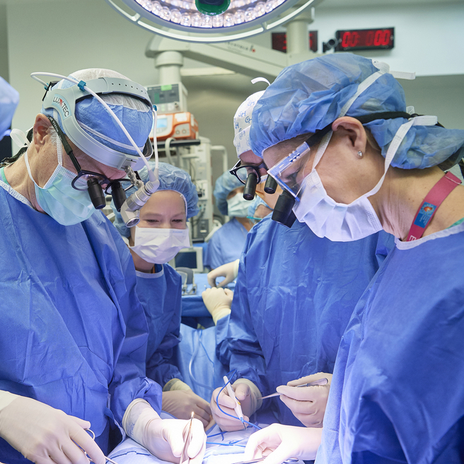
Rarely, an EXIT procedure may be required to deliver the baby. In an EXIT procedure, our SDU team will partially deliver the baby so that they are still attached to the placenta and receiving oxygen through the umbilical cord. This procedure allows time for fetal surgeons to establish an airway and remove the bronchogenic cyst. After the cyst is removed and your baby can breathe on their own, they will be fully delivered.
Your team will include a nurse coordinator, a maternal-fetal medicine specialist, and a pediatric and fetal surgeon. We will work with you to develop a treatment plan that is best for you and your family and is safe for the newborn.
Treatment for bronchogenic cyst
If the bronchogenic cyst is small and the baby is asymptomatic at birth, routine care is advised, including a CT scan at 1-2 months of age, with surgical resection of the cyst to follow. Surgical resection is generally recommended because of risk of recurrent lung infections later in life and development of cancer (very rare).
If the baby is symptomatic at birth, a chest X-ray and CT scan may be necessary as soon as possible, with surgical resection of the cyst sooner rather than later.
Long-term outlook
The vast majority of children with congenital lung lesions, including bronchogenic cyst, do extremely well and have normal lung function after their lesions are removed. This is thanks to rapid compensatory lung growth that occurs during childhood. Having surgery early maximizes this compensatory growth. No long-term follow up is required in these cases.
Children with moderate to large lesions can also do extremely well, but their outlook depends on expert treatment to avoid potential complications. These children may require long term follow-up and in rare cases support through our Pulmonary Hypoplasia Program (PHP).

Tour our Fetal Center
The Wood Center for Fetal Diagnosis and Treatment has cared for many families and will help you through your journey, too.

What to expect
From the moment of referral through delivery and postnatal care, your family can expect a supportive experience when you come to us with a diagnosis of a birth defect.
Resources to help
Bronchogenic Cyst Resources
Richard D. Wood Jr. Center for Fetal Diagnosis and Treatment Resources
Learning your baby has a birth defect is a life-changing experience. We want you to know that you are not alone. To help you find answers to your questions, we've created this list of educational health resources.
Meet your team
You will receive care from a highly skilled and compassionate team with a global reputation. This team helped shape modern fetal care and continues to improve techniques and treatments.