Fetal Echocardiogram
What is a fetal echocardiogram?
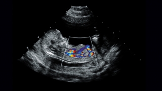 Fetal echocardiogram.
A fetal echocardiogram is a specialized ultrasound that provides a detailed view of your baby’s heart. Also known as a fetal echo, this test is used to examine the structure and function of your baby's heart before he or she is born.
Fetal echocardiogram.
A fetal echocardiogram is a specialized ultrasound that provides a detailed view of your baby’s heart. Also known as a fetal echo, this test is used to examine the structure and function of your baby's heart before he or she is born.
Fetal echocardiograms help us identify and diagnose congenital heart defects before birth. Our specially trained team performs an average of more than 3,700 fetal echo studies a year through our Fetal Heart Program, an unmatched level of experience.
We are also one of very view cardiac programs to offer early fetal cardiovascular imaging (EFCI), which allows us to diagnose heart problems as early as 12 weeks’ gestation. We use the most state-of-the-art technology in all of our exams, so you can be confident that you and your baby are getting the best care available.
Who you might meet
- Registered cardiac sonographer (RCS): A certified health professional that specializes in the use of ultrasound equipment and performs the fetal echocardiogram. Cardiac sonographers are technologists who use ultrasounds to examine the heart chambers, valves, and vessels. CHOP's sonographers are specially trained in scanning the fetal heart and have decades of fetal echocardiography experience. This dedicated team sets our ability to perform fetal echocardiograms apart from other centers.
- Pediatric cardiologist: A cardiologist is a physician with special training and skill in finding, treating and preventing diseases of the heart and blood vessels. Our cardiologists have advanced specialized training in evaluating and diagnosing fetal heart anomalies and conditions affecting fetal cardiovascular physiology.
What to expect
A warm gel is applied to your abdomen to help transmit sound waves to your uterus. You may be asked to move into different positions in order to optimize imaging of your baby’s anatomy.
A fetal echocardiogram procedure typically takes 45 to 60 minutes for a single gestation.
Private examination rooms have overhead monitors so you and your family can watch the ultrasound being performed. After the sonographer has completed the detailed fetal echocardiogram, they review the exam with the attending pediatric cardiologist.
If a heart problem is identified, you will meet with a pediatric cardiologist, a Fetal Heart Program nurse coordinator and a social worker to discuss the findings.