Reviewed on 07/22/2026
What is tracheoesophageal fistula?
Tracheoesophageal fistula, also referred to as TE fistula or TEF, is a rare condition that develops before a baby is born. The trachea is the windpipe that brings air from the throat to the lungs. In normal development, the trachea forms separately from the esophagus, or food pipe. A TEF is an abnormal connection in one or more places between the trachea and the esophagus. This can harm a baby’s breathing and can sometimes result in food or liquids being inhaled into the lungs.
TEF frequently occurs alongside esophageal atresia (EA), a condition in which the esophagus does not form a continuous connection to the stomach. Because these conditions affect both the airway and digestive tract, babies with EA/TEF require coordinated, highly specialized care shortly after birth.
How common is tracheoesophageal fistula?
Tracheoesophageal fistula (TEF) is a rare congenital condition, or birth defect, that occurs in approximately 1 in 3,500 to 4,500 babies. It commonly occurs with esophageal atresia (EA), a condition affecting the esophagus, and other congenital anomalies. It’s possible for TEF to occur in adults, but this is very rare.
What are the types of tracheoesophageal fistulas?
There are several known types of tracheoesophageal fistula. They’re generally classified by their association with or without esophageal atresia (EA).
TEF types include:
- Type A: This is a type of EA that is present without TEF. It’s usually classified as “long gap” and requires specialized repair (e.g., Foker process).
- Type B: This is a rare form of TEF that occurs with EA, on the upper part of the esophagus, and is also often classified as “long gap” EA.
- Type C: This is the most common type of TEF, accounting for roughly 85% of cases. It occurs with EA and affects the lower part of the esophagus.
- Type D: This very rare type of TEF that occurs with EA and impacts both the upper and lower parts of the esophagus.
Early evaluation of your baby’s TE fistula type is critical for determining the appropriate treatment plan.


What are the symptoms of tracheoesophageal fistula?
Symptoms of tracheoesophageal fistula (TEF) vary depending on whether it occurs with esophageal atresia (EA). Because TEF is rarely present without EA, the two conditions are often associated and share overlapping symptoms.
The most common EA and TEF symptoms include:
- Difficulty breathing
- Coughing or choking when eating
- Frothy, white bubbles coming from the mouth
- Unable to successfully feed by mouth
- Very round, full belly (from gas being trapped there)
- Bluish color to the skin, especially when the baby is eating
- Clinician is unable to pass a tube from the baby's mouth into their stomach
In the rare cases where TEF occurs without EA, it may take several weeks to diagnose the condition because symptoms usually only appear as mild coughing or respiratory problems. In these cases, babies are still able to successfully swallow and get food and liquid to their stomachs. However, the liquid can leak through the connection from the esophagus to the trachea and get into the lungs, causing infections or pneumonia.
What causes tracheoesophageal fistula?
Tracheoesophageal fistula (TEF) occurs during fetal development, when the trachea and esophagus fail to separate properly. It’s typically detected soon after birth and is commonly present with EA. While the direct cause of TEF is not yet known, research suggests there is likely a genetic component. This is because roughly half of babies born with TEF are also born with other health concerns such as VACTERL association, which is a group of conditions that occur together, impacting several organs and systems.
How is tracheoesophageal fistula diagnosed?
Most babies with tracheoesophageal fistula (TEF) also have esophageal atresia (EA) and are diagnosed shortly after birth, when symptoms first appear. However, when TEF occurs on its own, it may take weeks for symptoms to appear and be diagnosed.
Common TEF diagnosis methods include:
- Pediatric endoscopy: After a child is asleep by sedation or general anesthesia, an endoscope — a thin, flexible tube with a light and camera on the end — is inserted into the mouth to visualize any connections between the trachea and esophagus.
- Bronchoscopy: A small camera is put down the trachea to look for fistulas or connections that are small, hidden or not obvious from prior tests.
- X-rays with a nasogastric tube: A nasogastric (NG) tube is inserted through the baby’s nose into the esophagus, aiming for the stomach. If the tube meets a blockage, suggesting esophageal atresia (EA), X-rays can confirm the obstruction and its location. X-rays can also reveal gas in the stomach, indicating a connection between the lower esophagus and trachea.
- Pediatric ultrasound: A pediatric ultrasound uses sound waves to produce live images of the organs and soft tissues inside the body.
How is tracheoesophageal fistula treated?
Surgery is the primary treatment for repairing tracheoesophageal fistula (TEF). In some cases, multiple procedures may be needed. The type of surgery will depend on the following factors:
- The location of the TEF and the impact on the baby’s breathing
- The overall health of a baby, including any heart conditions or prematurity
When a fistula is detected between the esophagus and trachea, separating these tubes is usually the first step of surgery. At the time of surgery, your baby will be given a breathing tube, if they don’t already have one, to help keep their airway open during surgery. A pediatric anesthesiologist will then give them medicine to keep them comfortable for the operation.
Surgery is done through the side of the baby’s chest, using either one larger incision or several small ones, depending on the procedure.
After surgery, the baby returns to the N/IICU. Depending on the condition and the type of surgery performed, your baby may have an IV for pain medications, a G tube to carry food to the stomach, and/or a chest tube to drain fluid away from their reconnected esophagus. They may also need a ventilator to help them breathe until the child can breathe on their own.
In some cases, a small TEF may be treated endoscopically by a team of GI, pulmonary and ENT specialists. This may require multiple attempts overtime but could potentially remove the need for additional operations.
What are the risks of tracheoesophageal fistula repair?
The primary treatment for tracheoesophageal fistula (TEF) is surgery, which, like any procedure, involves potential risks. These risks vary based on the type of TEF your baby has and their overall health. Potential complications may include infection, narrowing of the esophagus known as an esophageal stricture, leakage, or recurrent fistula. Recurrent TE fistulas may occur months or even years after initial surgical repair. In our experience, one of the best ways to repair a recurrent TEF is to separate the healing esophagus from the healing trachea with a specialized surgery called posterior tracheopexy.
How long is the recovery process of tracheoesophageal fistula treatment?
Once your baby’s tracheoesophageal fistula (TEF) is repaired surgically, the most important next step is to allow time for healing. Each child’s treatment and recovery journey is unique, and at CHOP, our care teams are here to support every phase.
Some children will need just one surgery known as a “primary repair.” Some children may have planned staged surgeries, while others may have a need for future surgeries to improve the esophagus or trachea as they grow and develop.
The timing of a baby’s discharge after surgery will depend on many things, including:
- Whether they were born prematurely
- If the esophagus could be connected right away
- Respiratory status
- Whether other medical problems were identified
If your baby was born prematurely, they will likely need to remain in the hospital for several weeks or even months after surgery. Only a few weeks may be necessary if:
- Your baby was close to full term
- The esophagus could be connected right away
- There was not a leak after the esophagus was repaired
Before a child is discharged from the hospital, CHOP families are taught how to:
- Feed their baby if they have a feeding tube
- Give medications, if needed
- Mix formula or breast milk if increased calories are needed
- Take care of an incision and dressing
- Spot signs of possible respiratory infection
- Know when to call the pediatrician or surgeon with concerns
Following surgery, your baby will not be able to eat by mouth until they have healed. Some babies with TEF will require a surgical feeding tube (G tube) as they practice feeding by mouth. However, many children with an uncomplicated repair can learn to successfully eat by mouth without the aid of a G tube.
What is the prognosis of tracheoesophageal fistula?
Babies with tracheoesophageal fistula (TEF) who have no other anomalies and receive early treatment have a very high survival rate. Children with a history of TEF disease can thrive following surgical repair but may need ongoing monitoring from a dedicated healthcare team to keep an eye on growth, respiratory health, and potential long-term issues like feeding challenges or reflux.
Is tracheoesophageal fistula preventable?
Tracheoesophageal fistula (TEF) is a birth defect that forms before a baby is born and is generally not preventable due to its potential genetic factors. However, with early diagnosis and treatment, TEF can be repaired, and babies can go on to live normal, healthy lives.
CHOP’s areas of expertise and research for tracheoesophageal fistula
Through innovation in diagnostic imaging, advanced treatment methods, long-term management strategies and ongoing research to improve outcomes, CHOP leads the world in delivering care to children with congenital conditions like tracheoesophageal fistula (TEF).
While most babies with TEF are diagnosed shortly after birth, advances in prenatal imaging can detect symptoms before delivery. Findings such as excess amniotic fluid (polyhydramnios) or a small or absent stomach on high-resolution fetal ultrasound may prompt further evaluation.
When TEF is suspected before birth, families can benefit from early consultation with a multidisciplinary team. The Richard D. Wood Jr. Center for Fetal Diagnosis and Treatment at CHOP helps families plan for delivery, immediate breathing and feeding management, and surgical care after birth — helping reduce uncertainty and improve early outcomes.
Resources to help
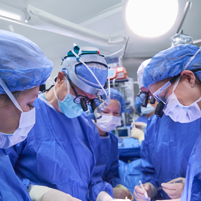
Meet your team
Your child will be cared for by a team with exceptional knowledge and experience in the care of these specific esophageal and airway conditions from before birth throughout childhood.