Ear, Nose and Throat Educational Illustrations
Dr. Brian Dunham is a surgeon and medical illustrator who specializes in the anatomy and diseases of the ear and skull base. He has published illustrations in multiple scientific articles and chapters and is actively involved in the development of novel applications of scientific art as an educational tool. The vast majority of his illustrations start as graphite drawings on paper, which are then scanned into a digital format and hand-rendered in Adobe Photoshop. He majored in Fine Arts as an undergraduate and pursued graduate level training in biomedical illustration at the Johns Hopkins Department of Art as Applied to Medicine. He is currently a professional member of the Association of Medical Illustrators and one of the founding members of Stream Studios at CHOP.
Anomalous facial nerve
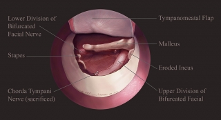 This Photoshop® rendering shows a bifurcated facial nerve as it courses through the middle ear space. This is a relatively rare finding. The illustration was developed from an intraoperative sketch drawn with a sterile skin marker on the sterile paper wrapper in which surgical gloves are packaged.
Facial nerve anatomy
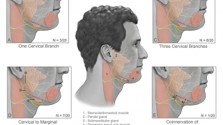
This composite Photoshop rendering describes the variable anatomy of the lower branches of the facial nerve found during a cadaveric study of the facial nerve. The facial nerve enables movement of the eyebrows, eye closure, and movement of the corners of the mouth. It courses from its intracranial origins through the internal auditory canal, into the middle ear and mastoid portion of the temporal bone (bony prominence behind the ear). It then exits the ear space anterior to the external ear and travels through the parotid gland to innervate muscles of the face. It is often at risk during both otologic and head & neck operations.
Laryngotracheoplasty
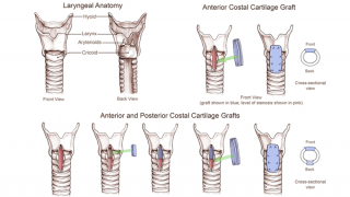 Composite illustration showing anterior and posterior graft laryngotracheoplasty for repair of subglottic stenosis, a condition in which the airway below the vocal cords narrows, often eventually producing respiratory distress. Prior to repair, many children with this condition have to breathe through a tracheostomy tube. Successful repair of their narrowed airway often allows them to forego the need for a tracheostomy tube.
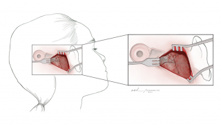
Transnasal transphenoidal approach to pituitary
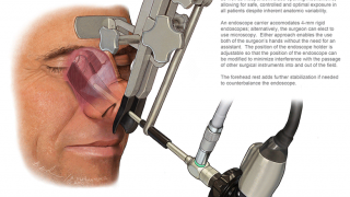 This composite Photoshop rendering details a transnasal approach to the pituitary gland with a novel retractor that was developed by Drs. Richard Chole and Ralph Dacey at Washington University. This particular approach and retractor allow for the use of rigid endoscopes and/or an operating microscope. It is an operation that otolaryngologists and neurosurgeons often perform jointly, predominantly in adults. Dr. Dunham worked on this illustration over a three-year period, as the retractor’s design was refined.
Cochlear implant fixation
This Photoshop illustration describes a technique for securing the receiver-stimulator of a cochlear implant to a patient’s skull using a resorbable plate and screws. The receiver-stimulator sits inside a soft tissue pocket created during the operation. As the wound heals, this soft tissue pocket eventually constricts around the receiver-stimulator and secures it to the skull. The plate and screws biodegrade over time and are fully resorbed within a year.
Human skull carbon dust rendering
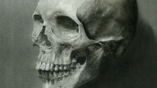
Carbon dust rendering of a human skull. Carbon dust is an older technique that was promoted by Max Brödel, considered by many to be the founding father of biomedical illustration in the United States. Tones are applied using dry sable brushes dipped in carbon dust as well as carbon pencils. The details produced with this technique can be very fine. This rendering was the first assignment that Dr. Dunham completed during his graduate level coursework. Being the only surgical trainee in his class, his teachers and mentors were kind enough to allow him to draw a skull; all the other students were asked to draw a hip bone.
Vestibular schwannoma - skull head composite
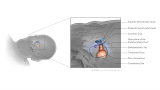
This illustration shows the relevant anatomical structure during a surgical approach to a vestibular schwannoma, a tumor that arises from the nerves that course inside the internal auditory canal. The patient is presented in surgical position. Some of the critical structures, namely the semicircular canals that are shown are covered in bone and not exposed during removal of the tumor. This illustration was created as a visual aid for a discussion on the limits of bony dissection during removal of these tumors.