Birth of a Breakthrough: Spina Bifida Video Series

The "Birth of a Breakthrough" video series explores spina bifida diagnosis, treatment options, delivery and follow-up care at The Children’s Hospital of Philadelphia. Experts at CHOP’s Center for Fetal Diagnosis and Treatment have the world’s greatest collective experience in prenatal repair for spina bifida. Hear from clinicians and patient families who have undergone fetal surgery for myelomeningocele, the most severe form of spina bifida.

En Español »

Transcript

Brief Introduction

Katherine Mulligan, mother: It was our first child. We were very excited, and of course, we were doing the countdown like every new parent does.

Michael Mulligan, father: We were looking forward to having our first ultrasound for our new baby and start our family together.

Katherine Mulligan, mother: "Well-baby ultrasound" they called it. You know, just to check everything out. No big deal. We weren't even going to find out if we were having a boy or a girl.

Michael Mulligan, father: And, of course, we didn't get exactly the great news we were expecting.

Katherine Mulligan, mother: No. The technician found that our child's brain was not forming correctly. So we had to go to a higher level ultrasound.

Michael Mulligan, father: So we did that and they confirmed that we had a baby with spina bifida.

Katherine Mulligan, mother: And, of course, our lives were turned upside down. All those hopes, dreams and everything else you had for your child were just gone.

N. Scott Adzick, MD: In the context of families getting a new diagnosis of spina bifida, they may or may not know much about it.

Mark P. Johnson, MD: It must not only be quite a shock, but because they may not understand what that means, it's probably pretty terrifying.

Leslie N. Sutton, MD: And we started talking about paralysis and catheters and shunts and wheelchairs, and they start to cry because this is not what they imagined.

Lori J. Howell, RN: All of their hopes and dreams for that child are readjusted, are reorganized.

Susan R. Miesnik, MSN: Most of the time they get this diagnosis before they actually come to see us, so they've had some time to adjust to it.

Lori J. Howell, RN: And one of our jobs at our center is to make sure that mothers have every option available to them.

N. Scott Adzick, MD: ...to educate them, to advise them, to help them so that whatever choice the mother and the family makes is the right one for them.

Spina Bifida Facts: What is Spina Bifida?

N. Scott Adzick, MD: Spina bifida or a more severe form, myelomeningocele, is the most common central nervous system or neurologic birth defect in the world.

Mark P. Johnson, MD: Myelomeningocele is a defect in the spine. It's failure of the spinal cord to close.

N. Scott Adzick, MD: The spinal cord, or the neural tube as the embryologists say, forms very early in gestation, certainly before 8 weeks' gestation. And spina bifida is thought to be a failure of the formation and closure of that neural tube. So that the spinal cord elements are exposed to the intrauterine environment.

Mark P. Johnson, MD: It's a progressive process. It's a progressive injury. So that by the end of pregnancy, there's been direct traumatic injury to the spinal cord, and there's been injury from the amniotic fluid itself.

Natalie E. Rintoul, MD: Because of changes in pressure and fluid flow, babies are also at risk for developing hydrocephalus.

N. Scott Adzick, MD: ...which is a fluid buildup on the brain and which damages the developing brain.

Leslie N. Sutton, MD: Virtually every child with myelomeningocele or spina bifida has an Arnold Chiari malformation on X-ray. And what it is, is that the back part of the brain called the "cerebellum" that has a lot to do with control of motor movements is wedged down into your neck.

N. Scott Adzick, MD: The cerebrospinal fluid that cushions the brain and cushions the spinal cord leaks out through the back through the open spina bifida defect. That leads to the back part of the brain getting sumped down into the upper part of the spinal canal. That, in turn, can block the circulation of cerebrospinal fluid in the brain because the fluid is produced in the brain and then there's no way for it to circulate beyond that blockage point.

Leslie N. Sutton, MD: So if there's a blockage in the flow of that fluid, then the water builds up inside the ventricle. So you've probably heard the term "water on the brain." The water isn't on the brain. It's inside the brain. If you don't treat that then you end up with the massively enlarged head. You die. Or you end up with severe mental retardation.

N. Scott Adzick, MD: So in order to minimize that ongoing damage of pressure from the fluid, the pediatric neurosurgeon places a shunt tube.

Leslie N. Sutton, MD: It's a simple device. This is a shunt. It's made out of silicone. This end goes through a hole in the back of the head, into that water cavity, and tunnels down under the skin, and goes into the abdomen which is able to absorb the fluid. It's not a cure. It treats the symptom. It doesn't treat the disease.

Natalie E. Rintoul, MD: Once you have a shunt there are multiple complications that can arise from it.

N. Scott Adzick, MD: The shunt can become infected, which is a serious complication because then the brain and the layers of the brain can become infected with meningitis encephalitis. And with each bout of infection, that can effect the brain function.

Leslie N. Sutton, MD: And then, of course, there are mechanical problems. They clog. They break. They get to be too short. They migrate.

N. Scott Adzick, MD: So it's not unusual for a child with spina bifida who has a shunt to require that shunt, and not only for life, but to have that shunt changed at least five or six times during the course of their life.

Mark P. Johnson, MD: It's important for parents to understand the potential complications or ramifications of spina bifida after the diagnosis is made.

Natalie E. Rintoul, MD: A child may experience difficulty with apnea, forgetting to breathe. They may also have breathing problems, choking, blue spells.

Leslie N. Sutton, MD: In the worst case, you end up with a child that's on a ventilator, with a tracheostomy, and a tube in the stomach to eat. And that is a huge impact on your quality of life.

Mark P. Johnson, MD: Spina bifida can cause paralysis in the lower extremities. That can interfere with the baby's ability to walk, or they may even be wheelchair dependent.

Natalie E. Rintoul, MD: They also have difficulty with their bowel and bladder function.

Leslie N. Sutton, MD: Most of these kids, when they become older, have to catheterize or else they have to wear diapers.

Mark P. Johnson, MD: They can develop significant curvatures of the spine because of instability — bony instabilities of the spine if they have a high lesion.

N. Scott Adzick, MD: Because of the lack of innervation to the feet and the ankles, there can be the formation of club feet, or the medical term is "talipes" as a consequence. So that requires orthopedic intervention after birth.

Leslie N. Sutton, MD: And then there are problems that can come on later on in life. It's not even necessarily done when you're a baby. You can get tethered cord, which means that the nerve tissue gets caught up in scar tissue, and you can lose function. So it's a lifelong issue of disability.

Evaluation: Learning Your Options

Lori J. Howell, RN: When a mom first calls our center, either our nurse coordinator or genetic counselor will talk with them about how they first learned about the diagnosis of spina bifida, what tests they've had so far, and their understanding of the diagnosis.

Susan R. Miesnik, MSN: They are scheduled for an entire day of diagnostic testing and counseling.

N. Scott Adzick, MD: The diagnostic process involves radiologic studies, maternal-fetal ultrasound that's usually 2 plus hours in duration, in which we look at everything.

Mark P. Johnson, MD: ...how big the lesion is, what segments of the spine are involved. They look for the degree of hydrocephaly, evidence for the Chiari malformation. And we look for evidence of paralysis or function of the lower extremities.

N. Scott Adzick, MD: Fetal MRI – that usually takes 30 - 45 minutes and requires the mother to be still inside the MRI chamber. A fetal echocardiogram to look at the fetal heart.

Jack Rychik, MD: Fetuses with myelomeningocele typically do not have any problems that are wrong with the fetal heart. However, as a fetal cardiologist, it's very important to make sure that we're not dealing with any abnormalities whatsoever of the heart, even if they're mild.

Mark P. Johnson, MD: Part of the evaluation when a family comes to our center is not only the detailed imaging of the fetus, but careful evaluation of the mother herself.

Lori J. Howell, RN: And then we meet with the family to go through the results of those tests and tell them what we know about the diagnosis.

N. Scott Adzick, MD: The counseling involves a multidisciplinary group – an experienced nurse, neurosurgeon, fetal surgeon, maternal-fetal medicine specialist, social worker. This is a team that can provide information to the family, review the results of the tests, and present the options.

Michael Mulligan, father: It was amazing how much information they were able to convey, and we understood it. Very caring and it just made you feel like, if they say this is an option for you, it was a potentially great thing for us.

Natalie E. Rintoul, MD: Families faced with the diagnosis of spina bifida in utero are now able to make choices.

Mark P. Johnson, MD: Many families, after hearing the range and potential severities of the disabilities and problems these children face, choose to terminate the pregnancy.

Leslie N. Sutton, MD: They have the option of staying here at CHOP, delivering here and we'll do conventional treatment, which is, we'll deliver the baby, and I'll close the back, put in the shunt, whatever's necessary in the newborn period.

Natalie E. Rintoul, MD: Or now they have the option of pursuing fetal surgery, which really does change the natural history of the disease as we knew it before.

Considering Fetal Surgery for Spina Bifida

N. Scott Adzick, MD: For the first time in the history of mankind outside of postnatal repair of myelomeningocele, we have a new way to offer hope to the family and to the child with prenatally diagnosed spina bifida.

Leslie N. Sutton, MD: Early studies that we did suggested that fetal surgery might be beneficial, but to prove it, we had to do a randomized prospective clinical trial.

N. Scott Adzick, MD: The MOMS Trial started in 2003, and it was a head-to-head comparison between repair of myelomeningocele after birth to repair of myelomeningocele before birth.

Leslie N. Sutton, MD: And what the MOMS Trial has basically showed us is that the fetal surgery group has done significantly better in virtually every way compared to conventional treatment.

Mark P. Johnson, MD: It's very important that the family understands that fetal surgery is a really, really, really big deal.

N. Scott Adzick, MD: The mother has to go through an operation. She's an innocent bystander in all this. She has to undergo a big operation to potentially benefit her fetus.

Mark P. Johnson, MD: It's a complicated multidisciplinary surgery. There are significant risks of the surgery itself.

Leslie N. Sutton, MD: We have to tell them that they could lose the baby. We could do fetal surgery, and they could wake up not being pregnant.

N. Scott Adzick, MD: There's a risk that's substantial for the fetus, in terms of the risk of premature birth. For the mother, there is a risk of just general anesthesia and a big operation.

Martha G. Hudson, MSW: For some families fetal surgery is not going to be an option medically for the mother or the child.

Susan R. Miesnik, MSN: If the fetus does not have hindbrain herniation, for example, an Arnold Chiari malformation, that fetus would not be eligible. If the lesion, the defect, is really low in the spine in the sacral area, a lot of times we don't offer that surgery, also.

N. Scott Adzick, MD: Sometimes, with regard to fetal intervention, the patients are referred too late. So we will not do the fetal operation after 26 weeks' gestation, for instance, because there appears to be no significant benefit.

Susan R. Miesnik, MSN: If the mother has certain medical conditions, it may exclude her from being offered this procedure – if the mother has severe diabetes or has severe asthma or has some issues with obesity or has mental health issues that are not well controlled. There are certain criteria that must be met. So the mother needs to be healthy in all aspects of her life.

Michael Mulligan, father: There are risks involved, but we knew that if they thought we were a good candidate, that it was potentially the right thing for us to do.

Katherine Mulligan, mother: A risk we were willing to take.

Michael Mulligan, father: Right.

Katherine Mulligan, mother: There is nothing more important than bringing that child into the world. And if we could get him here healthier, it meant the world to us.

Fetal Repair at CHOP: How It Works

Natalie E. Rintoul, MD: Once a family decides to pursue fetal surgery, things happen pretty rapidly.

N. Scott Adzick, MD: All the components of our rather large team meet to discuss the case and then have the parents come in, and we go through the entire process once again so that there's a clear understanding of what's involved and give them the opportunity to ask any additional questions.

Katherine Mulligan, mother: We had to be down here – I don't know if it was 5 o'clock or 6 o'clock, but it was very early in the morning. I was so nervous.

Michael Mulligan, father: And, you know, still a lot of consultations and things like that going on and, you know...

Katherine Mulligan, mother: Flurry of activity. They put an epidural in. It was the first thing they were going to do, and I had to be awake for that. You are knocked out for the surgery. And I remember bending over getting that epidural, and they're talking to me and all of a sudden, you go – Oh! I mean, if it hadn't hit us already, it hit me then: this is big time.

Michael Mulligan, father: Right.

Katherine Mulligan, mother: Seventeen days before that life was normal, and all of a sudden here we are in fetal surgery for something that's cutting edge.

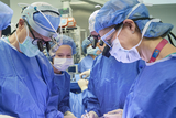
Leslie N. Sutton, MD: We have a special OR that we use for fetal surgery. It's a very large OR because there's a lot of equipment that's there, and there are a lot of people involved in this. There are a couple of anesthesiologists. There's the fetal team.

Susan R. Miesnik, MSN: Two to three surgeons in the room. There's a cardiologist in the room who monitors the baby's heart through the entire procedure. There's a neurosurgeon who actually does the myelomeningocele repair. There is an obstetrician or maternal-fetal medicine specialist in the room who monitors the mother's uterus and performs ultrasound so that we make sure we put the incision in the uterus where we need to put it.

Mark P. Johnson, MD: And it all has to be carefully choreographed, so that everybody is focused on what their role is, what they need to do, but everybody has to work seamlessly together.

N. Scott Adzick, MD: The mother goes under deep general anesthesia. Deep general anesthesia is important for at least three reasons. It anesthetizes the mother, it anesthetizes the fetus, and also gives us very good uterine relaxation — relaxation of the uterine muscle, which is very important for the operation. An ultrasound is done to look at the fetal position and then a sideways wound is made – almost hipbone to hipbone halfway between the belly button and the pubic bone to expose the uterus. Ofttimes the obstetrician will move the fetus within the uterus so that the myelomeningocele, or spina bifida lesion, is right underneath where we're going to make the cut in the uterus because the baby basically stays inside the uterus during the operation. The cut in the uterus is done again under ultrasound guidance – sterile, intraoperative, ultrasound guidance – with a uterine stapling device that fires absorbable staples that pinch off all the blood vessels and keep the membranes around the baby tacked up to the muscle layer of the uterus. During the operation the fetal cardiologist does monitoring of the fetal heart by echocardiography.

Jack Rychik, MD: We're able to look at blood flow patterns through the heart, the various structures, and the flow towards the different organs. Heart function. We look at the heart rate. Are able to feed that information back to the surgeons and to the anesthesiologist that's managing the mother.

N. Scott Adzick, MD: The neurosurgeon then comes in, and he and I do the myelomeningocele repair exactly like you would do in a newborn after birth.

Leslie N. Sutton, MD: I trim the sac of abnormal skin away from the nerve tissue. It's important to do that because that skin tissue, if I leave it behind, can become a cyst. It can be a problem later on. We then put the nerve tissue back into the spinal canal where it's supposed to be and then I try to get a three-layer closure. The first layer is the dura – that's the normal covering of the brain and the spinal cord. The second layer is the fascia – that's kind of like muscle-type tissue. And then the third layer is the skin.

N. Scott Adzick, MD: The goal is to cover the unprotected spinal cord so that it's a watertight protective closure. It needs to be watertight for two reasons. One, is that you want to stop the damage caused by amniotic fluid exposure for the rest of the pregnancy. And you also want to stop the leakage of cerebrospinal fluid out through the back that plays an important role in the development of hindbrain herniation and hydrocephalus. So if you stop the leak back here, the hindbrain can go back, and the hydrocephalus is less of an issue. Once the uterine wound is closed, the maternal abdomen is closed, including the skin, with a plastic surgical closure. All the layers are closed with an absorbable stitch that will dissolve once the mother's wound is healed.

Post Fetal Repair Surgery: The First Days After Surgery

Lori J. Howell, RN: For the first 24 to 48 hours after fetal surgery, really what we're looking at is fetal well-being and assessing for preterm labor.

Mark P. Johnson, MD: In the first few days, keeping the patient from going into preterm labor can be quite challenging. It requires use of several different medications.

Lori J. Howell, RN: Some of these medications have side effects which we need to explain to the parents before the surgery so that they know when they feel like a wet dishrag or their heart rate goes up a little bit or they feel a little weak, that that's a side effect we need to know about, and we can then change the medication accordingly.

Mark P. Johnson, MD: It's important to monitor the patient very carefully, so we do ultrasounds. We do fetal echocardiographic evaluations.

Jack Rychik, MD: We will perform fetal cardiovascular imaging on the day of the surgery itself, once mom has recovered and she's back in her room. And we'll do so for the first day, second and third day.

Martha G. Hudson, MSW: We certainly aim to get the mom out in four days, but we are pretty conservative, and if the mom is having any type of postsurgical issues, she will stay in the Hospital.

Mark P. Johnson, MD: If things go well, then the moms are discharged but have to remain at restricted activity for weeks after the surgery because of the risks of preterm labor or the risks of membrane separations that could lead to major complications.

Spina Bifida Care During Pregnancy

N. Scott Adzick, MD: One of the things that we've learned from open fetal surgery, in general, and for fetal surgery repair of myelomeningocele, specifically, is that it is an enormous commitment not only on behalf of our institution, our team, but most importantly, on behalf of the family.

Lori J. Howell, RN: When we talk to a mother and offer fetal surgery as an option, we need to know that they can comply with the travel arrangements, that they can stay with us so that we can monitor the pregnancy after fetal surgery.

Mark P. Johnson, MD: Weekly monitoring is very important to pick up any signs of complications following the surgery because identification of any problems will require an immediate change in the management plan.

Natalie E. Rintoul, MD: Families that relocate here from far away, they need to get other family members to take care of siblings and the like. It's a big – it's a big commitment.

Lori J. Howell, RN: It's really difficult, and some families just can't do it. And that's OK. And for those that opt for a fetal surgery intervention, we're here to provide that support.

Delivery After Fetal Surgery

Lori J. Howell, RN: Our goal with fetal surgery repair for spina bifida is to get the mother to 37 weeks.

N. Scott Adzick, MD: Babies who undergo fetal surgery tend to be born prematurely. In the MOMS trial they were born at an average gestational age just beyond 34 weeks, but 10 percent were born at 30 weeks or prior to that time, which is quite premature.

Mark P. Johnson, MD: Over 65 percent of them get past that 35-week mark. And then it becomes a matter of timing of delivery.

Susan R. Miesnik, MSN: We've made an incision in the uterus that is not in a safe part of the uterus to allow the woman to labor in any pregnancy going forward.

Lori J. Howell, RN: So the baby is delivered via cesarean section in the Special Delivery Unit.

N. Scott Adzick, MD: The Special Delivery Unit was built for the mother carrying a baby with a birth defect, was built for the mother carrying a baby with spina bifida.

Natalie E. Rintoul, MD: We, as the resuscitation team, feel like we are very prepared for those deliveries. We have excellent communication with the obstetricians, and we have access to all their prenatal imaging. We know those babies very well, and we coordinate their delivery and their care.

Katherine Mulligan, mother: Dr. Johnson delivered Sean.

Michael Mulligan, father: He came out very healthy, 6 lbs. 7 oz.

Katherine Mulligan, mother: Which was a very good size for a child that's born at 36 1/2 weeks. He was a big boy.

Susan R. Miesnik, MSN: We always make a point of bringing the baby to the mother prior to bringing the baby down to the NICU so that the mother and father – and any relatives that they want – can see the baby.

Natalie E. Rintoul, MD: And from there, we take the baby straight down to the newborn infant ICU and the moms are close. You know, they can come down as soon as they're feeling well. And, if there's something going on, we're able to go straight back up to their unit.

Michael Mulligan, father: The idea of being together is huge when you're talking about what's already a very stressful situation.

Natalie E. Rintoul, MD: So really the SDU has been a wonderful thing.

Another Choice: Postnatal Surgery for Spina Bifida

Mark P. Johnson, MD: It's important to understand that not all families want to have fetal surgery because of the requirements and the commitments and the risks that are involved.

Susan R. Miesnik, MSN: Families may come to us, undergo the diagnostic testing, and the defect may not be a defect that is included in the criteria for the surgery.

N. Scott Adzick, MD: Even if a family is an ideal surgery candidate, this may not be the best thing for them. And it is our obligation to help them understand that that's all right. That that's OK. That that's their choice. That that is the right choice for them.

Mark P. Johnson, MD: For families that want the traditional approach of having surgery after birth but still want to deliver at our center because of our multidisciplinary experience and care, we follow them as well. We, generally, have them come back at monthly intervals so we can monitor growth and development of baby – to look for their progression in the hydrocephaly and all the markers of complications. We, generally, deliver them at around 37 weeks.

N. Scott Adzick, MD: The mother, usually, has a cesarean section in the Special Delivery Unit.

Mark P. Johnson, MD: Our goal is to deliver the baby without any additional trauma to the myelomeningocele sac and also not to rupture the sac but to deliver the myelomeningocele intact. The baby then is evaluated by a neonatal resuscitation team.

Natalie E. Rintoul, MD: We have special equipment ready so that we can do different positions knowing that that spinal cord is still exposed.

Mark P. Johnson, MD: The baby then goes to the NICU where all the multiple subspecialties come and evaluate the baby. Neurosurgery, generally, take the baby to the operating room within the first 24 to 48 hours of life and do the neurosurgical repair.

Leslie N. Sutton, MD: The technical aspects are pretty much the same whether I do it prenatally or postnatally. I have the same layers. I do essentially the same operation. The beauty of our facility is that we have our own delivery suite right here in the Children's Hospital. So mother and baby are at the same place. So mother delivers. We get to look at the baby. We close the back the next day. I can talk to the mother about how things went, and she can hold the baby the next day.

Newborn Care at CHOP After Spina Bifida Surgery

Martha G. Hudson, MSW: In the postnatal course, regardless of whether or not you have had fetal surgery or postnatal surgery, our aim is get the mom down to the NICU as soon as possible to be with her baby. The babies all have the exact same workup regardless of which treatment they have had.

Lori J. Howell, RN: They are descended upon by a team of experts from urology, orthopedics, the spina bifida clinic, and a variety of folks from those areas to get the baseline tests that they might need.

Leslie N. Sutton, MD: We look at the head to see is there any sign that the water is building up with the hydrocephalus. We look at the legs and the feet to see if they're moving. And then we order certain radiographic studies, the MRIs, to look at those things from the inside.

Natalie E. Rintoul, MD: The near-term babies, 35 weeks and above, are often quite mature. I mean, they need a little bit of help feeding and maybe a little respiratory support, but we feel pretty good once they get to that gestation. And then kids between 26 to 32 weeks, they're young. We have to, you know, follow them very closely for all the regular, routine issues of prematurity.

N. Scott Adzick, MD: One of the important things in terms of neonatal care, particularly for premature babies, is a level of expertise that's unparalleled.

Natalie E. Rintoul, MD: The newborn infant ICU at the Children's Hospital of Philadelphia is one of the leading units in the country, if not the world. The unit has a lot of experience taking care of babies, preterm infants up to 6 months, up to a year. But what's very important for these families is that there are physicians and nurses, respiratory therapists, a whole team that know how to take care of a baby that's had fetal surgery.

N. Scott Adzick, MD: That is just not a baby who's born at 29 weeks gestation. That is a baby who's had open fetal surgery several weeks previously, born very premature at 29 weeks gestation. So you want expertise in that NICU that can care for any consequence for that baby, whether it's 2:00 in the morning or 2:00 in the afternoon, and that's the sort of expertise and depth of care that we have.

Michael Mulligan, father: It's tough to watch this happening to your loved ones, and you just feel much better knowing that there's this support staff. That they have an understanding of what consequences can come, of what outcomes could be. And they're ready for those outcomes.

Lori J. Howell, RN: I always tell parents that if your due date was April 1st and you're going to deliver at 37 weeks, that you'll probably go home around April 1st.

Natalie E. Rintoul, MD: So expect to be with us until your due date. Some kids graduate a little earlier, and some kids that may have had some complications may be with us a little longer. But that's a pretty good rule of thumb.

Martha G. Hudson, MSW: When a child is ready to go home from the NICU, we make sure they are completely hooked up in a spina bifida clinic in their area. If they are local, we will have them hooked up in our spina bifida clinic, and we will make sure that all the services they need in their community are set up prior to discharge.

Fetal Repair Follow-Up Care

Lori J. Howell, RN: Probably the most expectant time that families have is once they go home. What's going to happen then? And I think that is a major hallmark of our center that we continue to follow these babies.

N. Scott Adzick, MD: It's important to bring all the healthcare professionals to the baby. And so that's what we do in the multidisciplinary spina bifida clinic at CHOP.

Natalie E. Rintoul, MD: The spina bifida program at the Children's Hospital of Philadelphia was the first multidisciplinary program in the world.

N. Scott Adzick, MD: So there's 50 years plus of experience.

Natalie E. Rintoul, MD: It's an interdisciplinary comprehensive spina bifida program that really is geared at long-term management. And there are hundreds of kids in the program as well as adults.

N. Scott Adzick, MD: The follow-up of these babies is very important for their care. But one additional issue and benefit is that these patients teach us.

Lori J. Howell, RN: Only with that ongoing commitment of these parents to share their lives and their children with us are we going to continue to provide that information for future generations.

N. Scott Adzick, MD: There will be more lessons to learn that will enhance care, that will enhance counseling, and that will enhance the choices that families can make.

Superior Spina Bifida Treatment: Experience, Innovation, Excellence

Lori J. Howell, RN: For years, we have continued to focus and refocus all of our efforts on a baby with a birth defect. So when you come to CHOP and the Center for Fetal Diagnosis and Treatment, you have the benefit of all of that expertise.

Susan R. Miesnik, MSN: It's the same team. We've been working with the same people. We're like a finely oiled machine.

Natalie E. Rintoul, MD: So for a family who's carrying a fetus with spina bifida, you really want to go to a place that knows very well and has seen, you know, all the problems that can occur so that there really are no surprises.

Lori J. Howell, RN: Hearing your concerns and being able to answer those questions is incredibly reassuring to families.

Michael Mulligan, father: We'll all face challenges in life. But it's nice to start off without some of those major challenges when you can, you know, have science and medicine come together to make the life better.

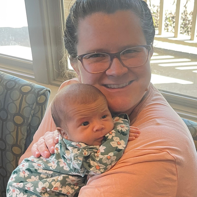
Katherine Mulligan, mother: Sean came out with his toes wiggling, kicking and screaming so to speak, and he's been doing that ever since. He runs. He jumps. He plays. He does everything he wants to do.

Michael Mulligan, father: He knows that there's some limitations, but I think he also knows that he's very fortunate. And we don't dwell on the spina bifida, but we don't ignore it either. It's just...

Katherine Mulligan, mother: It's part of our lives.

Michael Mulligan, father: Yeah, it's part of who Sean is. It's no question about that.

Katherine Mulligan, mother: We're very lucky that his quality of life, and ours, is just incredible, absolutely incredible.

Martha G. Hudson, MSW: It's wonderful that we can provide families with all these options and give the families the opportunities to learn everything they can about their babies.

N. Scott Adzick, MD: It takes a team. It takes a multidisciplinary team working in a very collegial, cooperative, fully integrated way.

Leslie N. Sutton, MD: You could not possibly do this surgery without this kind of a team.

N. Scott Adzick, MD: And that sort of experience is irreplaceable.

Leslie N. Sutton, MD: It is volume. It's having patients, steadily doing this, keeping the team sharp. That's critical to the success of this.

Natalie E. Rintoul, MD: It is easy to get excited and be committed when the entire team is committed.

Mark P. Johnson, MD: And I think that's what makes CHOP a really unique place. Very supportive of research. Very supportive of families. Very supportive and focused on doing whatever it takes to improve outcomes for children.

Topics Covered: Fetal Surgery for Spina Bifida (Myelomeningocele, MMC) Spina Bifida Causes, Symptoms and Treatment

Related Centers and Programs: Richard D. Wood Jr. Center for Fetal Diagnosis and Treatment


You Might Also Like
Connor selfie with parents and wooded valley behind him

A Star is Born

Doctors closed a hole in Connor's spine while he was still in his mother's womb. Follow his spina bifida journey.

Screen grab from Fetal Surgery for Spina Bifida video

Surgery Before Birth: Spina Bifida

This video about fetal surgery for spina bifida explains spina bifida treatment options and the benefits of spina bifida surgery before birth.

parents in Garbose SDU

Family First: Special Delivery Unit

Watch this video about CHOP's Special Delivery Unit, the first birthing unit of its kind for families expecting babies with a prenatally diagnosed birth defect.