MFM/Cardiology Collaboration Improves Fetal Management and Care
Published on
In Utero InsightsPublished on
In Utero InsightsFor more than 10 years, pediatric cardiologists from The Children’s Hospital of Philadelphia’s Fetal Heart Program (FHP) have offered state-of-the-art diagnostic, counseling and management services to fetuses with all forms of cardiovascular disease and their families. Through collaboration with the Center for Fetal Diagnosis and Treatment (CFDT), expertise from all facets of prenatal care is brought to bear on conditions affecting the fetal heart, ranging from the simple to the most complex, helping to ensure the best outcomes.
A one-day evaluation involves a series of cardiac and obstetrical tests unparalleled in scope. Early gestational fetal cardiovascular imaging (EFCI) is available for high-risk patients, and allows diagnosis of heart anomalies as early as 12 weeks’ gestation. Specially trained fetal sonographers and fetal cardiologists perform and interpret a fetal echocardiogram. High-level obstetrical ultrasound and ultra-fast fetal MRI add to the comprehensive understanding and provide context for many of the cardiovascular conditions seen. Information gathered from the evaluation is carefully reviewed by cardiologists and maternal-fetal medicine (MFM) specialists. After the evaluation, family counseling is provided by a dedicated team consisting of an FHP nurse coordinator, social worker, fetal cardiologist and MFM specialist. This integrated approach transcends traditional discipline perspectives and biases, and leads to an optimal evaluation focused on the needs of the fetus and family.
The FHP’s focus on the cardiovascular manifestations of congenital malformations that are not primary cardiac defects but may secondarily affect the heart reflects the unique collaborative philosophy that exists. For example, fetal cardiologists and MFM specialists combined their expertise to develop a cardiovascular scoring system for twin-twin transfusion syndrome (TTTS) that enables the team to not only discern which fetuses might benefit from fetal intervention, but also better predict which patients with TTTS might require further cardiac services after birth.*
The FHP’s expertise goes beyond diagnosis, as it leads the effort to attempt to treat life-threatening anomalies prior to birth. A team of fetal cardiologists, MFM specialists and cardiac catheterization experts has begun to offer fetal cardiac intervention in select cases. This team has successfully placed a stent across the atrial septum in a fetus with hypoplastic left heart syndrome and intact atrial septum. As a result, a child who more than likely would have died as a neonate is now a year and a half old and thriving.
“There is no one specialty that offers all of the expertise necessary to do this,” says Jack Rychik, MD, director of the FHP, one of the nation’s first dedicated programs focused on fetal cardiovascular care. “You have to have the hands-on skill to pass a needle into a target that is a foot away but only a fraction of an inch in diameter, and you need to have the ultrasound skills to be able to image all of this. But the ability to interpret these images before, during and after these procedures requires a deep understanding of cardiovascular physiology that only an experienced fetal cardiologist can bring to the table.”
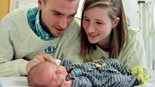 Tucker, the first survivor of fetal surgery for pericardial teratoma, pictured with his father Mike and mother Katie.
For rare cardiac conditions like pericardial teratoma, as well as for more common non-cardiac conditions like myelomeningocele, direct intraoperative imaging of the heart throughout open-fetal surgery is critically important. FHP cardiologists create a mini intensive care unit for the fetus and, through ultrasound and fetal echocardiography techniques, continuously monitor heart function, heart rate and flow patterns through the heart, providing ongoing, critically important feedback to the surgical team.
Tucker, the first survivor of fetal surgery for pericardial teratoma, pictured with his father Mike and mother Katie.
For rare cardiac conditions like pericardial teratoma, as well as for more common non-cardiac conditions like myelomeningocele, direct intraoperative imaging of the heart throughout open-fetal surgery is critically important. FHP cardiologists create a mini intensive care unit for the fetus and, through ultrasound and fetal echocardiography techniques, continuously monitor heart function, heart rate and flow patterns through the heart, providing ongoing, critically important feedback to the surgical team.
The team seamlessly transitions from fetal care to delivery and postnatal care. Depending on the condition, mothers may deliver in the Garbose Family Special Delivery Unit (SDU), the first birth facility in the world exclusively for mothers carrying babies with diagnosed birth defects. The SDU is equipped for immediate medical or surgical intervention upon delivery, when necessary. A delivery classification scale for cardiovascular conditions was specifically developed that allows the team to prepare for any problems a child may face upon delivery. For the most severe — who experience hemodynamic instability at separation from placental circulation — the team is prepared to perform an “immediate postpartum access to cardiac therapy” (IMPACT) procedure. After the mother undergoes a C-section in a cardiac operating room, the baby is whisked to an adjacent hybrid cath lab/OR suite for resuscitation. Cardiac catheterization to open blocked passages, heart surgery to repair defects, or pacing for heart block immediately after birth can be lifesaving, when seconds count.
“The resources necessary to prepare and organize this are tremendous,” Rychik says. “There’s no way you could gather all of these specialists and services at a moment’s notice. But by anticipating the needs of a fetus when separated from the mother, and properly classifying those who have the greatest need, we can achieve the best outcomes.”
To learn more about our fetal heart collaboration, please visit fetalsurgery.chop.edu/professional.
* Rychik J, Tian Z, Bebbington M, Xu F, McCann M, Mann S, Wilson RD, Johnson MP. The twin-twin transfusion syndrome: spectrum of cardiovascular abnormality and development of a cardiovascular score to assess severity of disease. Am J Obstet Gynecol. 2007 Oct;197(4):392.e1-8
Categories: In Utero Insights Summer 2013