Magnetoencephalography (MEG Scan)
What is a MEG scanner?
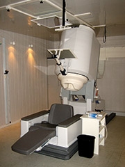 Magnetoencephalography (MEG) is an imaging technique that allows us to measure the magnetic fields produced by electrical activity in the brain.
The information from MEG assessments help us in a number of ways, including pinpointing the source of epilepsy when considering brain surgery; sensory mapping, identifying brain signatures associated with autism and helping researchers determine the function of various parts of the brain.
Advantages of MEG
- Noninvasive. With MEG, we can identify locations where seizures arise.
- Sensitive and accurate: MEG allows us to detect the tiny magnetic fields generated by brain activity and show where in the brain that occurs.
- Safe: MEG does not expose you or your child to radiation.
Advantages of MEG at CHOP
- Expertise: Our neuroradiologists are all board-certified experts in adult and pediatric neuroradiology. They have years of experience with MEG. All exams are read in conjunction with a PhD who specializes in MEG. Our team is one of the largest in the country.
- Care: Specially trained MEG technologists work with your family through the entire process, beginning with scheduling the MEG and helping with insurance pre-certification, performing a pre-MEG assessment and speaking with you and your child throughout the exam. A team of expert healthcare providers is also available if your child will be under anesthesia during the exam.
- Efficiency: with its two MEG machines, CHOP has one of the highest volume MEG centers in the world.
For more information, please call 267-426-7926 or 267-426-7927.
What should you do prior to the exam?
There are different preparations to follow according to the type of MEG scan your child is having. The technologist will call you to explain the preparations in detail prior to the exam.
If your child is:
- Being evaluated for epilepsy, your child cannot sleep the evening before the exam. If your child is taking medication, he should continue to do so. Do not stop meds! Your child will also have a brief MRI after the MEG scan. If a full clinical MRI is needed we will coordinate this with the MRI department.
- Being sedated for the exam, follow the General Anesthesia (GA) protocol in our sedation guidelines. All children up to 7 years old will undergo general anesthesia regardless of the reason for the exam. If your child is older and needs to be asleep for this procedure, general anesthesia is required because all other forms of sedation obscure seizure activity.
- Having any type of general motor or sensory mapping, no preparation is needed. Your child may sleep as usual the night before.
If your child has a noted allergy to radiographic contrast, additional preparations may be necessary.
Dress your child comfortably, in clothes that are easily removed (sweat clothes, t-shirts). Your child will be given a gown to change into for the MEG and MRI.
If you have copies of your child's previous imaging studies from another institution, please bring them for comparison.
MEG and MRI safety precautions
The magnetic field used to perform diagnostic images affects implanted medical devices and other items, such as braces. These will also affect the sensors of the MEG and MRI. For this reason, every patient must be screened to ensure both your and your child are safe to enter the magnetic field.
Note: Due to the sensitive nature of the MEG recording devices, parents or guardians will NOT be allowed to accompany their child into the MEG exam room.
Parents or guardians will be allowed to accompany their child into the MRI exam room. Other arrangements should be made for siblings.
Women who are pregnant or may be pregnant will be asked to leave the exam room during the procedure. Please make sure that there is someone else available to accompany your child during the exam, if needed.
What should you expect during the exam?
- If your child is receiving general anesthesia (GA), you will first be taken to our nursing station. Here, the GA team will place an IV in your child’s arm, hand or foot. Your child will feel only a little pinch.
- If your child is not receiving GA we will take you to the MEG scanner room. Our technologist will explain the procedure to both you and your child. Your child will be asked to change into a gown and lock up any necessary items. Parents may also use the locker.
- The technologist will position your child on the MEG table, most likely lying on his back, depending on the type of scan. The MEG scan requires us to place three leads on your child's head. We will also need to make three small dots with a skin marker in these areas.
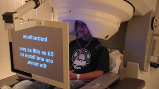
- We will dim the lights and explain to your child that we will be able to see and talk with him during the scan. Your child will then be moved into the MEG scanner helmet. The technologist will ask your child to hold very still during the scan. Any type of movement will make the data look blurry, and the scan may have to be repeated.
- We will begin the MEG scan when you and the technologist leave the room. The MEG scanner is very quiet. Your child will not feel any pain or discomfort associated with the MEG exam, but sometimes holding still for a long time can be uncomfortable.
- Your child may need additional scanning depending on his clinical history. If needed, we will move the bed into a seated position and ask your child to look at a video monitor and press buttons on a box. None of these devices cause any discomfort to your child.
- The data is collected on a computer screen and is sent to a team of physicians to be interpreted.
What should you do after the exam?
If your child received contrast for the MRI portion of the exam, be sure to give your child some extra liquids for the day.
If your child received general anesthesia, follow any instructions from the Anesthesia department.
Test results
The data from your child's exam must be processed and reviewed by several clinicians before a report is sent to your physician's office. This may take several weeks.
Your physician may call us at 215-590-2584 with questions regarding the report.
MEG case report
A typical case is that of a 9-year-old with medically refractory epilepsy for two years. His brain MRI was essentially normal and his doctors favored an anterior right temporal or frontal seizure onset, based on his seizure pattern and EEG findings.
MEG identified two independent areas from where seizures appeared to be arising, in the anterior and posterior aspects of the right temporal lobe.
Because of the MEG findings, the plan for the placement of subdural electrodes was significantly altered, as coverage was extended more posteriorly. Intracranial EEG confirmed the MEG findings and both regions were resected. The patient is now seizure free.