Pelvic Fracture Clinical Pathway — Emergency Department
Tips on Interpreting Pelvic Films
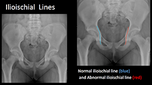
Ilioischial Line
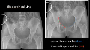
Iliopectineal Line
Pearls
Ilioischial Line: Radiographic landmark demarcating the POSTERIOR COLUMN of the acetabulum. Begins at the sciatic notch and extends inferiorly into the medial border of the ischium. Disruption of the ilioischial line reflects a posterior column acetabular fracture.
Iliopectineal Line: Radiographic landmark demarcating the ANTERIOR COLUMN of the acetabulum. Begins at the sciatic notch and extends along superior pubic ramus and symphysis pubis. Disruption of the iliopectineal line reflects an anterior column acetabular fracture.
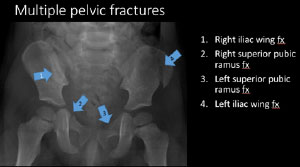
Multiple Pelvic Fractures
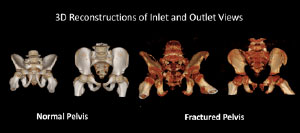
3D Reconstructions of Inlet and Outlet Views
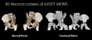
3D Reconstructions of Judet Views
Pearls
Computer tomography (CT) with 3D reconstructions allow for improved visualization, understanding, and classification of complex pelvic fractures, thus guiding therapeutic management. There are a variety of 3D surface-rendered reconstruction algorithms (maximum intensity projection, minimum intensity projection, etc.) that facilitates visual inspection of the bones or other anatomic structures.
Using 3D reconstructions, additional oblique radiographic images (Judet views) and caudal/cephalad radiographic images (inlet/outlet views) can be obviated.
Inlet view assess the pelvic ring integrity and potential ANTEROPOSTERIOR SHIFT (widening/narrowing) in pelvic fractures.
Outlet view assess VERTICAL SHIFT of the hemipelvis in pelvic fractures.
Judet views are used to evaluate the anterior column and the posterior column of the acetabulum using two oblique projections.