Deep Neck Space Infection Clinical Pathway — Emergency Department
Deep Neck Space Infection Clinical Pathway — Emergency Department
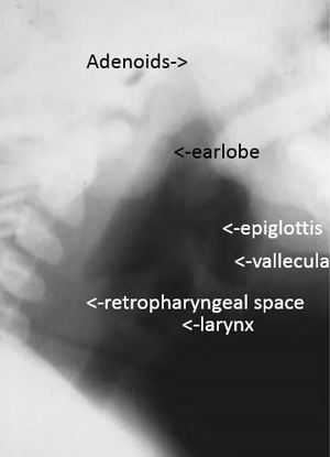
Interpreting Lateral Neck
Review the considerations and anatomic areas listed below when interpreting the lateral neck. A prevertebral space of < 6 mm at the level of C3 is considered normal in children. Alternatively, in general, in infants between 2 and 5 years of age, the retropharyngeal soft tissues (C1-C3) should be < 1/2 the AP length of C5 vertebral body, and the retrotracheal soft tissues (C4-C7) should be < the AP length of C5 vertebral body. In pediatric patients, widening of the prevertebral soft tissues can be a normal spurious finding that is related to expiration, crying, and/or suboptimal neck extension.
| Review the Following | Assessment |
|---|---|
|
|
|
|
|
|
|
|
|
|
|
|
|
|
|
|
|
|
Normal Upper Airway Anatomy—Infant
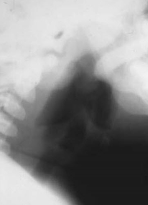
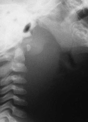
Retropharyngeal Cellulitis
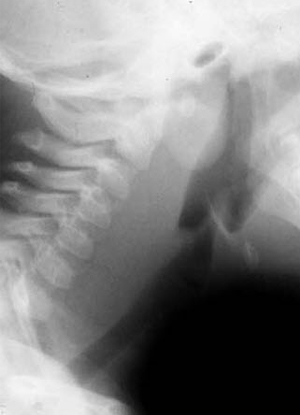
Retropharyngeal Abscess
Images courtesy of Richard Markowitz, MD