What is megaureter?
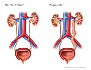
Megaureter is an abnormality of one or both of the ureters of a child. Ureters are the two funnel-shaped tubes that carry urine from the kidneys to the bladder. A megaureter refers to an expanded or widened ureter that does not function normally.
Whereas a normal ureter is about 3-5 mm, the size of a megaureter is usually greater than 7 mm in diameter.
Causes
A megaureter can be associated with the reverse flow of urine (vesicoureteral reflux, VUR). A megaureter can also be associated with an obstruction. The obstruction can either be the result of a ureterocele, or narrowing where the ureter meets the bladder (ureteral vesical junction obstruction, UVJO). The goal with megaureters is to determine which are obstructed, which have the reverse flow of urine, called reflux, and which have both.
A megaureter that is not associated with other problems occurs during fetal development. It occurs when a section of the ureter — which is normally a muscular layer of tissue — is replaced by stiff, fibrous tissue. Without the muscular layer of tissue, normal peristalsis (a worm-like movement of the ureter that propels urine toward the bladder) cannot occur.
Testing and diagnosis
The severity of the problem often determines how a diagnosis is made. Often a megaureter is diagnosed before birth by ultrasound. After birth, some children may have other problems that may suggest the presence of megaureter. This may prompt our team in the Division of Urology to perform further diagnostic tests, which may include the following:
- Renal bladder ultrasound: This procedure uses sound waves to outline the kidneys and bladder. If ureteral dilation is present, it will enable us to see the degree of dilation. It can also detect the presence of a ureterocele.
- Voiding cystourethrogram (VCUG): A catheter is placed through your child’s urethra into the bladder. The tube will be used to slowly fill the bladder with a solution. While the bladder is being filled, a special machine (fluoroscopy) is used to take pictures. The radiologist looks to see if any of the solution is going back up into the kidneys, which confirms the diagnosis of VUR.
- MAG III renal scan: This study will be performed to determine how each kidney is functioning and will determine the degree of blockage, if any. An intravenous line (IV) is used to inject a special solution called an isotope into the veins. The isotope makes it possible to see the kidneys clearly. Pictures of the kidneys will be taken will a large X-ray machine that rotates around your child.
- MRI/MRU: MRI is a radiation-free diagnostic procedure that uses a combination of a large magnet, radiofrequencies and a computer to produce detailed images of the body. Magnetic resonance urography (MRU) creates detailed pictures of the kidneys, ureters and bladder.
Treatment
Through research in the Division of Urology, we have shown that as long as kidney function is not significantly affected and urinary tract infections do not become an issue, some megaureters can be managed medically. (Journal of Urology, Prenatally Detected PMU, 2005. Read the abstract.) This may involve the use of antibiotic prophylaxis and radiology observation with repeated ultrasounds.
When the dilation is severe without showing signs of improvement, or kidney function is affected, surgical repair may be necessary.
Surgical options
Cutaneous distal ureterostomy: This may be necessary in a newborn with massive ureteral dilation or poor renal function. The ureter is surgically brought to the surface of the skin to allow it to drain urine freely into the diaper. This allows the affected kidney and ureter to decompress. Around 18 months of age, the ureter is then reimplanted into the bladder.
Ureterovesical junction obstruction: This surgical procedure involves removing the section of the ureter that is abnormal, reducing it and reconnecting the ureter. The segments of most megaureters regain tone once they are unobstructed.
Reviewed by: Division of Urology
Date: May 2011
Resources to help
Division of Urology Resources
Caring for a child with an illness or injury can be overwhelming. We have resources to help you find answers to your questions and feel confident in the care you are providing your child.

Meet your team
At CHOP, your child's care is in the hands of one of the largest and most accomplished teams of pediatric urology experts in the world.