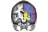
An international team including researchers from the Lifespan Brain Institute (LiBI) of Children’s Hospital of Philadelphia (CHOP) and the University of Pennsylvania have created a new tool that benchmarks brain development over the human lifespan, based on magnetic resonance imaging (MRI) data from more than 100,000 individuals. The work was jointly led by LiBI researchers and colleagues at the University of Cambridge. Described today in Nature, the interactive open resource, known as BrainChart, harmonizes brain images in a way that will allow researchers to measure brain development against reference charts like those used for evaluating children’s height and weight.
“There are no standardized growth charts for brain development like there are for other growth metrics such as height and weight, despite the fact that we know the brain goes through many changes over the human lifespan,” said co-senior author Aaron Alexander-Bloch, MD, PhD, Director of the Brain-Gene-Development Lab within the Department of Child and Adolescent Psychiatry and Behavioral Sciences at Children’s Hospital of Philadelphia and Assistant Professor of Psychiatry at the University of Pennsylvania. “Our work brings together a huge amount of imaging data that will continue to grow, allowing researchers and eventually clinicians to evaluate brain development against standardized measures.”
Growth charts that track age-related changes are a cornerstone of pediatric health care, but they only exist for height, weight, and head circumference for the first decade of life. The researchers recognized a need for tools that would allow for a systematic evaluation of brain development and aging, given that experts have learned that many psychiatric disorders are the result of atypical brain development. Neurodegenerative diseases like Alzheimer’s and Parkinson’s Disease also cause observable changes in the brain, and developmental genetic disorders lead to structural brain abnormalities. Given that mental illness and dementia collectively represent the single biggest global health burden, the researchers saw a need for a standardized chart that would serve as a benchmark for brain structure over the human lifespan.
To create the BrainChart platform, the researchers aggregated the largest neuroimaging dataset to date, consisting of 123,984 MRI scans from 101,457 participants ranging in age from 115 days after conception to 100 years old, drawn from more than 100 primary research studies. The researchers analyzed tissue volumes and other MRI metrics and applied centile scores and rates of change over the lifespan. By doing so, the paper identified previously unreported neurodevelopmental milestones, including an early growth period that starts around 17 weeks after conception, when the brain is 10% its maximum size, until 3 years of age, when the brain is approximately 80% its maximum size. The researchers also demonstrated that their centile scores, an important benchmark of any growth chart, were stable over time for individual assessments.
Importantly, by using a statistical framework that accounted for MRI data coming from different sources, times, and clinical conditions, the researchers were able to harmonize the data on a single platform, creating a common language that could be used to understand brain images from different sources. By standardizing the images in the dataset, all of which were acquired through prior research studies, the researchers also laid the groundwork for adding images acquired through routine scans in the clinic.
The researchers noted that although BrainChart could someday be useful in clinical settings, more research is needed to validate and refine the model so that it could be useful in diagnosing brain-related conditions. The immediate use is more likely to apply to researchers, who now have a resource that can provide a benchmark against which they can apply their own findings. Additionally, the research team emphasized that brain charts must be representative of the whole population, pointing to the need for more brain MRI data on previously under-represented socio-economic and ethnic groups.
“Just as there are still significant caveats when it comes to the diagnostic interpretation of where a child falls on a height, weight, and BMI chart, we would likewise expect that there will be nuances to how BrainChart could be used in a clinical setting,” said Jakob Seidlitz, PhD, co-first author of the study and a post-doctoral research associate working in Dr. Alexander-Bloch’s lab. “That said, by creating this common language for brain images, we have built the necessary bridge that will help bring imaging insights into clinical practice, which bodes well for future progress towards digital diagnosis of atypical brain structure and development.”
Together with Dr. Alexander-Bloch and Dr. Seidlitz, the work was led by Richard Bethlehem, PhD, Simon White, PhD, and Ed Bullmore, MB, PhD at the University of Cambridge.
The work was supported with funding from the National Institute of Mental Health (T32MH019112-29 and K08MH120564), the British Academy, the Autism Research Trust, UKRI Medical Research Council (MC_UU_00002/2), the NIHR Cambridge Biomedical Research Centre (BRC-1215-20014), the Medical Research Council, a NIHR Senior Investigator award, and the Wellcome Trust.
RAI Bethlehem et al. “Brain charts for the human lifespan,” Nature, April 6, 2022, DOI: 10.1038/s41586-022-04554-y
Featured in this article
Specialties & Programs
An international team including researchers from the Lifespan Brain Institute (LiBI) of Children’s Hospital of Philadelphia (CHOP) and the University of Pennsylvania have created a new tool that benchmarks brain development over the human lifespan, based on magnetic resonance imaging (MRI) data from more than 100,000 individuals. The work was jointly led by LiBI researchers and colleagues at the University of Cambridge. Described today in Nature, the interactive open resource, known as BrainChart, harmonizes brain images in a way that will allow researchers to measure brain development against reference charts like those used for evaluating children’s height and weight.
“There are no standardized growth charts for brain development like there are for other growth metrics such as height and weight, despite the fact that we know the brain goes through many changes over the human lifespan,” said co-senior author Aaron Alexander-Bloch, MD, PhD, Director of the Brain-Gene-Development Lab within the Department of Child and Adolescent Psychiatry and Behavioral Sciences at Children’s Hospital of Philadelphia and Assistant Professor of Psychiatry at the University of Pennsylvania. “Our work brings together a huge amount of imaging data that will continue to grow, allowing researchers and eventually clinicians to evaluate brain development against standardized measures.”
Growth charts that track age-related changes are a cornerstone of pediatric health care, but they only exist for height, weight, and head circumference for the first decade of life. The researchers recognized a need for tools that would allow for a systematic evaluation of brain development and aging, given that experts have learned that many psychiatric disorders are the result of atypical brain development. Neurodegenerative diseases like Alzheimer’s and Parkinson’s Disease also cause observable changes in the brain, and developmental genetic disorders lead to structural brain abnormalities. Given that mental illness and dementia collectively represent the single biggest global health burden, the researchers saw a need for a standardized chart that would serve as a benchmark for brain structure over the human lifespan.
To create the BrainChart platform, the researchers aggregated the largest neuroimaging dataset to date, consisting of 123,984 MRI scans from 101,457 participants ranging in age from 115 days after conception to 100 years old, drawn from more than 100 primary research studies. The researchers analyzed tissue volumes and other MRI metrics and applied centile scores and rates of change over the lifespan. By doing so, the paper identified previously unreported neurodevelopmental milestones, including an early growth period that starts around 17 weeks after conception, when the brain is 10% its maximum size, until 3 years of age, when the brain is approximately 80% its maximum size. The researchers also demonstrated that their centile scores, an important benchmark of any growth chart, were stable over time for individual assessments.
Importantly, by using a statistical framework that accounted for MRI data coming from different sources, times, and clinical conditions, the researchers were able to harmonize the data on a single platform, creating a common language that could be used to understand brain images from different sources. By standardizing the images in the dataset, all of which were acquired through prior research studies, the researchers also laid the groundwork for adding images acquired through routine scans in the clinic.
The researchers noted that although BrainChart could someday be useful in clinical settings, more research is needed to validate and refine the model so that it could be useful in diagnosing brain-related conditions. The immediate use is more likely to apply to researchers, who now have a resource that can provide a benchmark against which they can apply their own findings. Additionally, the research team emphasized that brain charts must be representative of the whole population, pointing to the need for more brain MRI data on previously under-represented socio-economic and ethnic groups.
“Just as there are still significant caveats when it comes to the diagnostic interpretation of where a child falls on a height, weight, and BMI chart, we would likewise expect that there will be nuances to how BrainChart could be used in a clinical setting,” said Jakob Seidlitz, PhD, co-first author of the study and a post-doctoral research associate working in Dr. Alexander-Bloch’s lab. “That said, by creating this common language for brain images, we have built the necessary bridge that will help bring imaging insights into clinical practice, which bodes well for future progress towards digital diagnosis of atypical brain structure and development.”
Together with Dr. Alexander-Bloch and Dr. Seidlitz, the work was led by Richard Bethlehem, PhD, Simon White, PhD, and Ed Bullmore, MB, PhD at the University of Cambridge.
The work was supported with funding from the National Institute of Mental Health (T32MH019112-29 and K08MH120564), the British Academy, the Autism Research Trust, UKRI Medical Research Council (MC_UU_00002/2), the NIHR Cambridge Biomedical Research Centre (BRC-1215-20014), the Medical Research Council, a NIHR Senior Investigator award, and the Wellcome Trust.
RAI Bethlehem et al. “Brain charts for the human lifespan,” Nature, April 6, 2022, DOI: 10.1038/s41586-022-04554-y
Contact us
Child and Adolescent Psychiatry and Behavioral Sciences