
Members of our fetal radiology team recently published a study on prenatal ultrasound (US) features of lung lesions that had a final surgical pathologic diagnosis of congenital lobar overinflation (CLO). To our knowledge, this study reports the largest number of surgically resected and pathologically proven cases of CLO, none of which had prenatal US or MRI features to suggest an obstructing cyst, mass or bronchial atresia as the obstructive etiology.
This study also describes a new US feature of CLO, specifically, hypervascularity defined as exuberant flow beyond the central portion of the lung that persists at high pulse repetition frequencies. The study also details CLO’s prenatal imaging course.
About congenital lobar emphysema
CLO, also known as congenital lobar emphysema, is a relatively rare pulmonary abnormality affecting approximately one in 20,000 to 30,000 births. Bronchial obstruction results in lobar or segmental alveolar hyperexpansion without underlying parenchymal maldevelopment. The hyperexpanded lung frequently causes compression of the adjacent normal lung and most commonly presents in the neonatal period with clinical signs of tachypnea or respiratory distress.
About the study
The researchers performed radiology and clinical database searches from 2001 to 2017 to identify prenatally diagnosed lung lesions that had a final diagnosis of CLO. All patients underwent multidisciplinary assessment and care by a team consisting of maternal-fetal medicine specialists, radiologists, cardiologists, geneticists and pediatric surgeons. Size, echotexture and vascularity were assessed with detailed US, and the signal characteristics and vascularity were assessed with fetal MRI. Follow-up prenatal US scans, postnatal imaging and postnatal outcomes were reviewed.
The study population consisted of 12 patients. The median gestational age was 23.3 weeks at the time of evaluation, and the median congenital pulmonary airway malformation volume-to-head circumference ratio was 0.66. Lesion location varied with six in the lower lobes (four right and two left), five in the upper lobes (three left and two right), and one in the right middle lobe. Lesion echotexture was homogeneously echogenic relative to the normal lung in all cases and none demonstrated macrocysts. Hypervascularity by color Doppler US was observed in five cases (41.7 percent). A T2-hyperintense lung lesion was identified by MRI in all 12 cases, with elongated vessels identified in 11 cases (91.7 percent). All 12 cases had pathologically proven CLO.
Study findings
In our researchers’ experience, the presence of hypervascularity in a solid echogenic lung lesion has proven to be the most salient finding in predicting CLO. Recognition of this US finding resulted in CLO being the favored prenatal diagnosis in the three most recent cases of CLO evaluated at our center. Given that CLO and microcystic CPAMs share other US features, it is reasonable to include the latter in the differential diagnosis of noncystic, hyperechoic/hyperintense lung lesions. In the absence of hypervascularity, there were no other US findings that were helpful in predicting CLO. The prenatal course detailed in the study also provides a better understanding that can aid in the counseling of prenatally suspected cases of CLO.
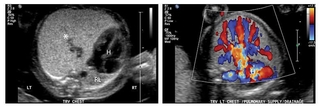
Transverse ultrasound image (left) demonstrates markedly increased echogenicity of the abnormal left lung (asterisk) relative to the compressed right lung (RL) with severe rightward shift of the heart (H). Color Doppler ultrasound (right) reveals marked hypervascularity of the expanded left lung with settings optimized for high flow (pulse repetition frequency, 1800 Hz).
Reference
Oliver, ER, DeBari SE, Horii SC, Pogoriler JE, Victoria T, Khalek N, Howell LJ, Adzick NS, Coleman BG. Congenital lobar overinflation: A rare enigmatic lung lesion on prenatal ultrasound and magnetic resonance imaging. J Ultrasound Med. 2018 Sep 12. [Epub ahead of print]
Featured in this article
Specialties & Programs
Members of our fetal radiology team recently published a study on prenatal ultrasound (US) features of lung lesions that had a final surgical pathologic diagnosis of congenital lobar overinflation (CLO). To our knowledge, this study reports the largest number of surgically resected and pathologically proven cases of CLO, none of which had prenatal US or MRI features to suggest an obstructing cyst, mass or bronchial atresia as the obstructive etiology.
This study also describes a new US feature of CLO, specifically, hypervascularity defined as exuberant flow beyond the central portion of the lung that persists at high pulse repetition frequencies. The study also details CLO’s prenatal imaging course.
About congenital lobar emphysema
CLO, also known as congenital lobar emphysema, is a relatively rare pulmonary abnormality affecting approximately one in 20,000 to 30,000 births. Bronchial obstruction results in lobar or segmental alveolar hyperexpansion without underlying parenchymal maldevelopment. The hyperexpanded lung frequently causes compression of the adjacent normal lung and most commonly presents in the neonatal period with clinical signs of tachypnea or respiratory distress.
About the study
The researchers performed radiology and clinical database searches from 2001 to 2017 to identify prenatally diagnosed lung lesions that had a final diagnosis of CLO. All patients underwent multidisciplinary assessment and care by a team consisting of maternal-fetal medicine specialists, radiologists, cardiologists, geneticists and pediatric surgeons. Size, echotexture and vascularity were assessed with detailed US, and the signal characteristics and vascularity were assessed with fetal MRI. Follow-up prenatal US scans, postnatal imaging and postnatal outcomes were reviewed.
The study population consisted of 12 patients. The median gestational age was 23.3 weeks at the time of evaluation, and the median congenital pulmonary airway malformation volume-to-head circumference ratio was 0.66. Lesion location varied with six in the lower lobes (four right and two left), five in the upper lobes (three left and two right), and one in the right middle lobe. Lesion echotexture was homogeneously echogenic relative to the normal lung in all cases and none demonstrated macrocysts. Hypervascularity by color Doppler US was observed in five cases (41.7 percent). A T2-hyperintense lung lesion was identified by MRI in all 12 cases, with elongated vessels identified in 11 cases (91.7 percent). All 12 cases had pathologically proven CLO.
Study findings
In our researchers’ experience, the presence of hypervascularity in a solid echogenic lung lesion has proven to be the most salient finding in predicting CLO. Recognition of this US finding resulted in CLO being the favored prenatal diagnosis in the three most recent cases of CLO evaluated at our center. Given that CLO and microcystic CPAMs share other US features, it is reasonable to include the latter in the differential diagnosis of noncystic, hyperechoic/hyperintense lung lesions. In the absence of hypervascularity, there were no other US findings that were helpful in predicting CLO. The prenatal course detailed in the study also provides a better understanding that can aid in the counseling of prenatally suspected cases of CLO.
Transverse ultrasound image (left) demonstrates markedly increased echogenicity of the abnormal left lung (asterisk) relative to the compressed right lung (RL) with severe rightward shift of the heart (H). Color Doppler ultrasound (right) reveals marked hypervascularity of the expanded left lung with settings optimized for high flow (pulse repetition frequency, 1800 Hz).
Reference
Oliver, ER, DeBari SE, Horii SC, Pogoriler JE, Victoria T, Khalek N, Howell LJ, Adzick NS, Coleman BG. Congenital lobar overinflation: A rare enigmatic lung lesion on prenatal ultrasound and magnetic resonance imaging. J Ultrasound Med. 2018 Sep 12. [Epub ahead of print]
Contact us
Richard D. Wood Jr. Center for Fetal Diagnosis and Treatment