
The Cardiac Center’s 3D Imaging Review Suite harnesses new technologies to deliver individualized, patient-specific care
Virtual reality — once the domain of gamers — now has new applications in pediatric cardiology, and the Cardiac Center at Children’s Hospital of Philadelphia (CHOP) is leading the exploration of this technology’s potential.
In the Cardiac Center’s 3D Imaging Review Suite, the result of an innovative collaboration between diagnostic imaging experts, cardiac clinicians take images from sources such as 3D echocardiograms, CTs and MRIs and review these images through virtual reality (VR) software known as SlicerVR. The result? Realistic, 3D models that clinicians can resize, interact with and even step into.
“You feel like you’re looking at something that’s right in front of you,” says Matt Jolley, MD, cardiac anesthesiologist and SlicerVR developer. “You can hold the image in your hand, you can move it, you can step into a ventricle and look around.”
With funding from the Cardiac Center, clinicians like Jolley are leveraging the capabilities of VR to educate surgical trainees, develop novel interventions and deliver highly-individualized, precision care.
Mapping surgical procedures
Surgeons use 3D imaging technology — such as 3D PDFs and printed models — to carefully plan interventions and optimize patient care. In the 3D Imaging Review Suite, clinicians can review a patient’s anatomy in multiple modalities, including VR, ensuring they have all of the information necessary to make clinical decisions about a patient’s care.
“We’ve gone from 3D technology being a helpful modality to being a routine clinical tool,” says Reena Ghosh, MD, who runs the 3D Imaging Review Suite with cardiac MRI attending Kevin Whitehead, MD, PhD. “Now a surgeon can walk through a patient’s heart before a case and figure out the best approach for that child.”
A Cardiac Center clinician reviews a patient’s heart anatomy in virtual reality.
In some cases, clinicians find that viewing these images through VR gives them access to hidden information not otherwise available. Jonathan M. Chen, MD, Co-Executive director of the Cardiac Center, says this is particularly the case when planning the repair of complex ventricular septal defects (VSD). A VSD — or “hole in the heart” — is an opening in the tissue between the heart’s lower chambers. A patient with complex VSDs, however, may have multiple openings in the heart’s tissue. The true size and locations of these openings may not be apparent on a 2D scan, such as a CT. VSDs are sometimes repaired through a minimally-invasive transcatheter procedure, but surgery can be needed. In order to plan the appropriate intervention in a patient with multiple VSDs, a cardiac clinician needs to know exactly where the openings are.
By viewing a patient’s scans in VR, a surgeon is able to see the heart anatomy exactly as it is, from multiple angles and perspectives, prior to surgery. This eliminates surprises and prevents additional, unexpected procedures. Says Dr. Chen, “There’s nothing else that gives you that kind of GPS.”
Another primary clinical use of VR is in planning the placement of intracardiac baffles — or tunnel connections — through which surgeons direct blood flow in complicated reconstructive heart surgeries, such as a biventricular repair. Through the use of VR and a technique called digital subtraction, a surgeon is able to identify the best pathways for these tunnels without the time or cost associated with 3D printing.
Virtual device selection and development
In the Pediatric Heart Valve Center at CHOP, virtual reality is also being used to inform device selection for transcatheter procedures. Many commercial valves are designed for adults and aren’t small enough for pediatric patients, and for children with rare heart disease, sometimes don’t exist at all.
Using SlicerVR technology and scans of a patient’s heart anatomy, cardiac interventionalists can virtually “test” the size and placement of existing devices to determine which one will best repair the defect and help avoid unforeseen consequences. For patients with rare heart disease, this software also enables clinicians to develop and test virtual prototypes of novel devices.
A Cardiac Center clinician uses VR to simulate device selection and placement.
“No one valve and no one kid are the same,” says Dr. Chen. “This is patient-specific, individualized medicine.”
Education and data sharing
Virtual reality also has capabilities in the education of residents, fellows and surgical trainees. The current Cardiac Registry is a large collection of actual heart specimens with different types of heart disease. Because some of these hearts are old and can be easily damaged, Lindsay S. Rogers, MD, attending cardiologist and Director of Education for the Echocardiography Laboratory at CHOP, is leading a project to increase their accessibility through VR.
Through a technique called photogrammetry, hundreds of pictures from multiple angles are taken of each heart and used to create a 3D image. The image of this specimen can then be viewed and interacted with in virtual reality.
An annotated heart specimen is explored in virtual reality.
“This is a great way to preserve our collection,” says Dr. Rogers. “We can now create novel educational content with the hearts that we have.”
This digitized registry, which includes hearts with rare conditions, can also be shared across institutions, benefiting pediatric heart patients all over the world.
The future of precision medicine
From pre-surgical planning to data sharing, the highly-advanced technology in the Cardiac Center’s 3D Imaging Suite is opening new doors in patient-specific medicine.
While other 3D technologies — such as 3D PDFs or printed models — are often the tools of choice for surgical procedures involving the spatial relationships of extracardiac vessels, VR provides a detailed image of the heart’s internal anatomy while also being less costly and more time efficient than other modalities. The Cardiac Center is currently conducting research that compares virtual reality to other 3D technologies with the goal of determining which technology is most beneficial for different types of heart disease.
In addition, researchers in the Pediatric Heart Valve Center hope to leverage VR capabilities in the development of a curated database that may one day inform valve repairs in patients with rare heart defects.
“Every kid is unique,” says Dr. Jolley. “Their images are unique. If you distill what we’re doing here, it’s image-based precision medicine.”
Featured in this article
Specialties & Programs
The Cardiac Center’s 3D Imaging Review Suite harnesses new technologies to deliver individualized, patient-specific care
Virtual reality — once the domain of gamers — now has new applications in pediatric cardiology, and the Cardiac Center at Children’s Hospital of Philadelphia (CHOP) is leading the exploration of this technology’s potential.
In the Cardiac Center’s 3D Imaging Review Suite, the result of an innovative collaboration between diagnostic imaging experts, cardiac clinicians take images from sources such as 3D echocardiograms, CTs and MRIs and review these images through virtual reality (VR) software known as SlicerVR. The result? Realistic, 3D models that clinicians can resize, interact with and even step into.
“You feel like you’re looking at something that’s right in front of you,” says Matt Jolley, MD, cardiac anesthesiologist and SlicerVR developer. “You can hold the image in your hand, you can move it, you can step into a ventricle and look around.”
With funding from the Cardiac Center, clinicians like Jolley are leveraging the capabilities of VR to educate surgical trainees, develop novel interventions and deliver highly-individualized, precision care.
Mapping surgical procedures
Surgeons use 3D imaging technology — such as 3D PDFs and printed models — to carefully plan interventions and optimize patient care. In the 3D Imaging Review Suite, clinicians can review a patient’s anatomy in multiple modalities, including VR, ensuring they have all of the information necessary to make clinical decisions about a patient’s care.
“We’ve gone from 3D technology being a helpful modality to being a routine clinical tool,” says Reena Ghosh, MD, who runs the 3D Imaging Review Suite with cardiac MRI attending Kevin Whitehead, MD, PhD. “Now a surgeon can walk through a patient’s heart before a case and figure out the best approach for that child.”
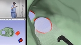
A Cardiac Center clinician reviews a patient’s heart anatomy in virtual reality.
In some cases, clinicians find that viewing these images through VR gives them access to hidden information not otherwise available. Jonathan M. Chen, MD, Co-Executive director of the Cardiac Center, says this is particularly the case when planning the repair of complex ventricular septal defects (VSD). A VSD — or “hole in the heart” — is an opening in the tissue between the heart’s lower chambers. A patient with complex VSDs, however, may have multiple openings in the heart’s tissue. The true size and locations of these openings may not be apparent on a 2D scan, such as a CT. VSDs are sometimes repaired through a minimally-invasive transcatheter procedure, but surgery can be needed. In order to plan the appropriate intervention in a patient with multiple VSDs, a cardiac clinician needs to know exactly where the openings are.
By viewing a patient’s scans in VR, a surgeon is able to see the heart anatomy exactly as it is, from multiple angles and perspectives, prior to surgery. This eliminates surprises and prevents additional, unexpected procedures. Says Dr. Chen, “There’s nothing else that gives you that kind of GPS.”
Another primary clinical use of VR is in planning the placement of intracardiac baffles — or tunnel connections — through which surgeons direct blood flow in complicated reconstructive heart surgeries, such as a biventricular repair. Through the use of VR and a technique called digital subtraction, a surgeon is able to identify the best pathways for these tunnels without the time or cost associated with 3D printing.
Virtual device selection and development
In the Pediatric Heart Valve Center at CHOP, virtual reality is also being used to inform device selection for transcatheter procedures. Many commercial valves are designed for adults and aren’t small enough for pediatric patients, and for children with rare heart disease, sometimes don’t exist at all.
Using SlicerVR technology and scans of a patient’s heart anatomy, cardiac interventionalists can virtually “test” the size and placement of existing devices to determine which one will best repair the defect and help avoid unforeseen consequences. For patients with rare heart disease, this software also enables clinicians to develop and test virtual prototypes of novel devices.
A Cardiac Center clinician uses VR to simulate device selection and placement.
“No one valve and no one kid are the same,” says Dr. Chen. “This is patient-specific, individualized medicine.”
Education and data sharing
Virtual reality also has capabilities in the education of residents, fellows and surgical trainees. The current Cardiac Registry is a large collection of actual heart specimens with different types of heart disease. Because some of these hearts are old and can be easily damaged, Lindsay S. Rogers, MD, attending cardiologist and Director of Education for the Echocardiography Laboratory at CHOP, is leading a project to increase their accessibility through VR.
Through a technique called photogrammetry, hundreds of pictures from multiple angles are taken of each heart and used to create a 3D image. The image of this specimen can then be viewed and interacted with in virtual reality.
An annotated heart specimen is explored in virtual reality.
“This is a great way to preserve our collection,” says Dr. Rogers. “We can now create novel educational content with the hearts that we have.”
This digitized registry, which includes hearts with rare conditions, can also be shared across institutions, benefiting pediatric heart patients all over the world.
The future of precision medicine
From pre-surgical planning to data sharing, the highly-advanced technology in the Cardiac Center’s 3D Imaging Suite is opening new doors in patient-specific medicine.
While other 3D technologies — such as 3D PDFs or printed models — are often the tools of choice for surgical procedures involving the spatial relationships of extracardiac vessels, VR provides a detailed image of the heart’s internal anatomy while also being less costly and more time efficient than other modalities. The Cardiac Center is currently conducting research that compares virtual reality to other 3D technologies with the goal of determining which technology is most beneficial for different types of heart disease.
In addition, researchers in the Pediatric Heart Valve Center hope to leverage VR capabilities in the development of a curated database that may one day inform valve repairs in patients with rare heart defects.
“Every kid is unique,” says Dr. Jolley. “Their images are unique. If you distill what we’re doing here, it’s image-based precision medicine.”
Contact us
Cardiac Center