Researchers at Children’s Hospital of Philadelphia (CHOP) found that the artificial intelligence (AI) chatbot, CHATGPT-4, showed promise as a viable tool to enhance ultrasound image analysis, effectively differentiating liver pathologies. The findings were recently reported in the journal JMIR AI.
The integration of AI into medical imaging, particularly with large language models like ChatGPT-4, has the potential to advance diagnostic workflows by enabling automated and rapid analysis. In this study, researchers aimed to evaluate ChatGPT-4's performance in liver ultrasound radiomics, assessing its ability to differentiate liver conditions – specifically fibrosis, steatosis, and normal liver tissue – compared with traditional analysis software.

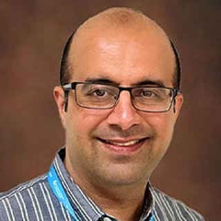
“With further refinement, ChatGPT-4 can be an effective and scalable tool to enhance patient outcomes in medical imaging,” said Laith Sultan, MD, MPH, a research scientist and principal investigator with the Center for Pediatric Contrast Ultrasound at CHOP whose research focuses on advanced ultrasound imaging techniques.
In the study, researchers analyzed 70 grayscale ultrasound images from a preclinical liver disease model – including groups with fibrosis, fatty liver and normal liver. They processed the dataset of liver ultrasound images through ChatGPT-4 and conventional radiomics methods, evaluating the performance of each method in terms of accuracy, efficiency and practicality in clinical settings.
The results showed that ChatGPT-4 performed comparably to traditional radiomics techniques in analyzing liver ultrasound images, achieving 76% accuracy and 83% sensitivity in distinguishing liver pathologies. While the traditional software achieved a slightly higher sensitivity of 89%, ChatGPT-4 added efficiency by processing multiple images concurrently.
Importantly, researchers noted that ChatGPT-4 has limitations. For example, the AI model’s interpretations are based on the data it was trained on, which may not encompass all possible variations in liver pathology. Therefore, researchers envision ChatGPT-4 as assisting rather replacing image analysis.
The project was supported by the CHOP Department of Radiology under the supervision of Hansel J. Otero, MD, Sudha Anupindi, MD, and Kassa Darge, MD, PhD.
Sultan et al. “ChatGPT-4-Driven Liver Ultrasound Radiomics Analysis: Advantages and Drawbacks Compared to Traditional Techniques.” JMIR AI. Online May 19, 2025. DOI:10.2196/68144.
Featured in this article
Experts
Specialties & Programs
Researchers at Children’s Hospital of Philadelphia (CHOP) found that the artificial intelligence (AI) chatbot, CHATGPT-4, showed promise as a viable tool to enhance ultrasound image analysis, effectively differentiating liver pathologies. The findings were recently reported in the journal JMIR AI.
The integration of AI into medical imaging, particularly with large language models like ChatGPT-4, has the potential to advance diagnostic workflows by enabling automated and rapid analysis. In this study, researchers aimed to evaluate ChatGPT-4's performance in liver ultrasound radiomics, assessing its ability to differentiate liver conditions – specifically fibrosis, steatosis, and normal liver tissue – compared with traditional analysis software.

“With further refinement, ChatGPT-4 can be an effective and scalable tool to enhance patient outcomes in medical imaging,” said Laith Sultan, MD, MPH, a research scientist and principal investigator with the Center for Pediatric Contrast Ultrasound at CHOP whose research focuses on advanced ultrasound imaging techniques.
In the study, researchers analyzed 70 grayscale ultrasound images from a preclinical liver disease model – including groups with fibrosis, fatty liver and normal liver. They processed the dataset of liver ultrasound images through ChatGPT-4 and conventional radiomics methods, evaluating the performance of each method in terms of accuracy, efficiency and practicality in clinical settings.
The results showed that ChatGPT-4 performed comparably to traditional radiomics techniques in analyzing liver ultrasound images, achieving 76% accuracy and 83% sensitivity in distinguishing liver pathologies. While the traditional software achieved a slightly higher sensitivity of 89%, ChatGPT-4 added efficiency by processing multiple images concurrently.
Importantly, researchers noted that ChatGPT-4 has limitations. For example, the AI model’s interpretations are based on the data it was trained on, which may not encompass all possible variations in liver pathology. Therefore, researchers envision ChatGPT-4 as assisting rather replacing image analysis.
The project was supported by the CHOP Department of Radiology under the supervision of Hansel J. Otero, MD, Sudha Anupindi, MD, and Kassa Darge, MD, PhD.
Sultan et al. “ChatGPT-4-Driven Liver Ultrasound Radiomics Analysis: Advantages and Drawbacks Compared to Traditional Techniques.” JMIR AI. Online May 19, 2025. DOI:10.2196/68144.
Contact us
Kaitlyn Tivenan
Center for Pediatric Contrast Ultrasound