Case
A 5-day-old infant was referred to the Emergency Department from her primary care doctor with tachypnea, a heart murmur, and decreased oral intake. She was born via cesarean section due to a breech presentation following an uncomplicated pregnancy. She was briefly in the NICU for respiratory distress, but was discharged home on day of life 3.
In the emergency department her labs were notable for hypoglycemia but were otherwise reassuring. Blood, urine, and cerebrospinal fluid cultures were sent, and she was started on antibiotics. A chest X-ray showed cardiomegaly and concern for pulmonary vascular congestion. An echocardiogram demonstrated 2 small ventricular septal defects but was otherwise structurally and functionally normal. She was started on a nasal cannula for tachypnea and was admitted to the neonatal intensive care unit for continued evaluation and management.
Discussion
During the first few days of hospitalization, this infant remained tachypneic, requiring high flow nasal cannula. While not clear on prior X-rays, a repeat chest X-ray was concerning for a possible right-sided diaphragmatic hernia versus diaphragmatic eventration. Ultrasound showed an intact right hemidiaphragm more consistent with diaphragmatic eventration. A CT chest showed marked anterior elevation of the right medial hemidiaphragm without clear herniation, also most consistent with a diaphragmatic eventration. She was then brought to the operating room by surgeon William Peranteau, MD, who found that a small piece of liver had herniated through a small anterior diaphragmatic defect. He was then able to reduce the hernia and repair the defect with a transversus abdominus muscle flap.
Congenital diaphragmatic hernia (CDH) occurs in up to 1 in 2 500 live births when the abdominal viscera herniate into the chest through a diaphragmatic defect. While prenatal referral to specialized high-volume centers, advanced ventilation strategies and pulmonary hypertension management, and extracorporeal membrane oxygenation (ECMO) have improved outcomes, mortality from CDH remains high between 25% to 30%. Given the unique and complex issues associated with CDH, infants with CDH benefit from experienced comprehensive multidisciplinary teams such as our Pulmonary Hypoplasia Program at Children’s Hospital of Philadelphia (CHOP).
Over 60% of CDH cases are diagnosed on a routine 18-to-22-week anatomy ultrasound. However, some cases may not be identified until later in pregnancy due to lack of early herniation of the abdominal contents into the fetal thorax, which can happen with a small defect, or because of technical or interpretative issues on an earlier exam. In more mild cases, a CDH may not be identified until postnatal life, either in the delivery room, or sometimes weeks, months, or even years later when the patient presents with mild gastrointestinal or respiratory symptoms or has a chest X-ray.
CDH vs. Diaphragmatic Eventration
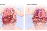
The most common type of CDH is a posterolateral hernia of the diaphragm, also known as a Bochdalek hernia, which accounts for approximately 80% to 90% of all CDH. About 85% occur on the left side, about 10% on the right, and approximately 5% are bilateral (see Figure 1). A posterolateral hernia is often accompanied by herniation of the stomach, intestines, liver, and/or spleen into the chest cavity.
Non-posterolateral (non-Bochdalek) hernias are anterior defects that can occur in the midline or on the right or left sides. These are more likely to be missed on prenatal imaging and often have a more mild presentation.
Finally, infants can have diaphragmatic eventration, which is incomplete muscularization of the diaphragm, as was initially suspected in our patient, Severe cases of diaphragmatic eventration can be associated with pulmonary hypoplasia and respiratory distress while milder degrees can present later in life with subtle respiratory symptoms.
Our patient had this less common anterior form of CDH, which likely accounts for her delayed diagnosis. Although a rare diagnosis, infants with ongoing respiratory distress and tachypnea should have an X-ray with careful attention to the possibility of a CDH.
She had an uneventful postoperative recovery and was slowly weaned off the ventilator and then extubated about 1 week postoperatively. She was discharged home on postoperative day 16 taking oral feeds and breathing comfortably without respiratory support. She will continue to follow with Dr. Peranteau as well as with CHOP’s multidisciplinary Pulmonary Hypoplasia Program long-term follow-up clinic.
References and Recommended Readings
Langham MR, Kays DW, Ledbetter DJ, et al. Congenital diaphragmatic hernia. Epidemiology and outcome. Clin Perinatol. 1996;23(4):671-688.
Shanmugam H, Brunelli L, Botto LD, Krikov S, et al. Epidemiology and prognosis of congenital diaphragmatic hernia: a population-based cohort study in Utah. Birth Defects Res. 2017;109:1451-1459.
Gupta VS, Harting MT, Lally PA, et al. Mortality in congenital diaphragmatic hernia: a multicenter registry study of over 5000 patients over 25 years. Ann Surg. 2023;1;277(3):520-527.
Hedrick HL, Adzick NS. Congenital diaphragmatic hernia in the neonate. In: UpToDate, Garcia-Prats (Ed), UptoDate, Waltham, MA, 2023.
Deprest J, Brady P, Nicolaides K, et al. Prenatal management of the fetus with isolated congenital diaphragmatic hernia in the era of the TOTAL trial. Semin Fetal Neonatal Med. 2014;19(6):338-348.
Al-Salem AH, Nawaz A, Matta H, et al. Herniation through the foramen of Morgagni: early diagnosis and treatment. Pediatric Surgery International. 2002;18(2-3):93-97
Featured in this article
Specialties & Programs
Case
A 5-day-old infant was referred to the Emergency Department from her primary care doctor with tachypnea, a heart murmur, and decreased oral intake. She was born via cesarean section due to a breech presentation following an uncomplicated pregnancy. She was briefly in the NICU for respiratory distress, but was discharged home on day of life 3.
In the emergency department her labs were notable for hypoglycemia but were otherwise reassuring. Blood, urine, and cerebrospinal fluid cultures were sent, and she was started on antibiotics. A chest X-ray showed cardiomegaly and concern for pulmonary vascular congestion. An echocardiogram demonstrated 2 small ventricular septal defects but was otherwise structurally and functionally normal. She was started on a nasal cannula for tachypnea and was admitted to the neonatal intensive care unit for continued evaluation and management.
Discussion
During the first few days of hospitalization, this infant remained tachypneic, requiring high flow nasal cannula. While not clear on prior X-rays, a repeat chest X-ray was concerning for a possible right-sided diaphragmatic hernia versus diaphragmatic eventration. Ultrasound showed an intact right hemidiaphragm more consistent with diaphragmatic eventration. A CT chest showed marked anterior elevation of the right medial hemidiaphragm without clear herniation, also most consistent with a diaphragmatic eventration. She was then brought to the operating room by surgeon William Peranteau, MD, who found that a small piece of liver had herniated through a small anterior diaphragmatic defect. He was then able to reduce the hernia and repair the defect with a transversus abdominus muscle flap.
Congenital diaphragmatic hernia (CDH) occurs in up to 1 in 2 500 live births when the abdominal viscera herniate into the chest through a diaphragmatic defect. While prenatal referral to specialized high-volume centers, advanced ventilation strategies and pulmonary hypertension management, and extracorporeal membrane oxygenation (ECMO) have improved outcomes, mortality from CDH remains high between 25% to 30%. Given the unique and complex issues associated with CDH, infants with CDH benefit from experienced comprehensive multidisciplinary teams such as our Pulmonary Hypoplasia Program at Children’s Hospital of Philadelphia (CHOP).
Over 60% of CDH cases are diagnosed on a routine 18-to-22-week anatomy ultrasound. However, some cases may not be identified until later in pregnancy due to lack of early herniation of the abdominal contents into the fetal thorax, which can happen with a small defect, or because of technical or interpretative issues on an earlier exam. In more mild cases, a CDH may not be identified until postnatal life, either in the delivery room, or sometimes weeks, months, or even years later when the patient presents with mild gastrointestinal or respiratory symptoms or has a chest X-ray.
CDH vs. Diaphragmatic Eventration
The most common type of CDH is a posterolateral hernia of the diaphragm, also known as a Bochdalek hernia, which accounts for approximately 80% to 90% of all CDH. About 85% occur on the left side, about 10% on the right, and approximately 5% are bilateral (see Figure 1). A posterolateral hernia is often accompanied by herniation of the stomach, intestines, liver, and/or spleen into the chest cavity.
Non-posterolateral (non-Bochdalek) hernias are anterior defects that can occur in the midline or on the right or left sides. These are more likely to be missed on prenatal imaging and often have a more mild presentation.
Finally, infants can have diaphragmatic eventration, which is incomplete muscularization of the diaphragm, as was initially suspected in our patient, Severe cases of diaphragmatic eventration can be associated with pulmonary hypoplasia and respiratory distress while milder degrees can present later in life with subtle respiratory symptoms.
Our patient had this less common anterior form of CDH, which likely accounts for her delayed diagnosis. Although a rare diagnosis, infants with ongoing respiratory distress and tachypnea should have an X-ray with careful attention to the possibility of a CDH.
She had an uneventful postoperative recovery and was slowly weaned off the ventilator and then extubated about 1 week postoperatively. She was discharged home on postoperative day 16 taking oral feeds and breathing comfortably without respiratory support. She will continue to follow with Dr. Peranteau as well as with CHOP’s multidisciplinary Pulmonary Hypoplasia Program long-term follow-up clinic.
References and Recommended Readings
Langham MR, Kays DW, Ledbetter DJ, et al. Congenital diaphragmatic hernia. Epidemiology and outcome. Clin Perinatol. 1996;23(4):671-688.
Shanmugam H, Brunelli L, Botto LD, Krikov S, et al. Epidemiology and prognosis of congenital diaphragmatic hernia: a population-based cohort study in Utah. Birth Defects Res. 2017;109:1451-1459.
Gupta VS, Harting MT, Lally PA, et al. Mortality in congenital diaphragmatic hernia: a multicenter registry study of over 5000 patients over 25 years. Ann Surg. 2023;1;277(3):520-527.
Hedrick HL, Adzick NS. Congenital diaphragmatic hernia in the neonate. In: UpToDate, Garcia-Prats (Ed), UptoDate, Waltham, MA, 2023.
Deprest J, Brady P, Nicolaides K, et al. Prenatal management of the fetus with isolated congenital diaphragmatic hernia in the era of the TOTAL trial. Semin Fetal Neonatal Med. 2014;19(6):338-348.
Al-Salem AH, Nawaz A, Matta H, et al. Herniation through the foramen of Morgagni: early diagnosis and treatment. Pediatric Surgery International. 2002;18(2-3):93-97
Contact us
Division of Neonatology