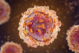
A growing body of literature has shown that mitochondria – the energy powerhouses of our cells – can be shared between different types of cells through miniscule tunnels, allowing mitochondria from one cell to travel to another. Studies have shown that these portals, called tunneling nanotubes, allow mitochondria to transfer from immune cells to cancer cells. Specifically, under tissue culture conditions, “spider-like” cancer cells are surrounded by a web of nanotubes that entangle nearby T cells to drain their mitochondria.
This phenomenon has raised numerous questions within the scientific community. How do these nanotubes form? What genes and pathways cause this transfer from immune cells to cancer cells, and why does this transfer only happen in one direction – from T cells to cancer cells – and not the other way around? Answering these questions would ideally involve studying this mitochondrial transfer under physiological conditions, but the current method to track mitochondria involves on genetic engineering of photo-activatable mitochondria and thus isn’t easily done in humans.
To circumvent this challenge, researchers from Children’s Hospital of Philadelphia (CHOP) have developed an algorithm called MERCI (Mitochondrial-Enabled Reconstruction of Cellular Interactions), which uses single cell RNA sequencing data and computational tools to determine which cells in patient tumor samples receive mitochondria from T cells. They derived this tool after observing and quantifying mitochondrial transfer in immune cells that were cultured alongside cancer cells, using single cell technology to identify which types of cancer cells were receiving mitochondria from T cells. Importantly, this work confirmed the unidirectional mitochondrial transfer at single cell genomic levels for the first time, allowing systematic investigation of this process.
After validating the computational tool, the researchers applied MERCI to human cancer samples and uncovered a previously unreported cancer phenotype related to mitochondrial transfer. The cancer cells engaged in mitochondrial transfer displayed higher cytoskeleton remodeling activity and elevated gene expressions in the tumor necrosis factor ɑ pathway. Using MERCI, the researchers identified 17 genes implicated in nanotube formation and mitochondrial transfer and created a gene set enrichment score – called a tumor MT transfer (TMT) score – based on the mRNA expression of these 17 genes. They then analyzed a large human patient cohort using TMT scores and found higher TMT scores correlated with poorer patient outcomes, likely due to T cell exhaustion and the promotion of cancer cell proliferation.
“MERCI is a powerful tool that could help gauge patient prognosis, as we have shown that higher TMT scores are associated with poorer outcomes in cancer patients,” said Bo Li, PhD, an investigator in the Center for Computational and Genomic Medicine and associate professor in the Department of Pathology and Laboratory Medicine at CHOP. “Future research should seek to identify which genes and pathways are most involved in this process, so that we can use this data to develop targeted treatments.”
Read more about this study, which was recently published in the journal Cancer Cell.
Featured in this article
Specialties & Programs
A growing body of literature has shown that mitochondria – the energy powerhouses of our cells – can be shared between different types of cells through miniscule tunnels, allowing mitochondria from one cell to travel to another. Studies have shown that these portals, called tunneling nanotubes, allow mitochondria to transfer from immune cells to cancer cells. Specifically, under tissue culture conditions, “spider-like” cancer cells are surrounded by a web of nanotubes that entangle nearby T cells to drain their mitochondria.
This phenomenon has raised numerous questions within the scientific community. How do these nanotubes form? What genes and pathways cause this transfer from immune cells to cancer cells, and why does this transfer only happen in one direction – from T cells to cancer cells – and not the other way around? Answering these questions would ideally involve studying this mitochondrial transfer under physiological conditions, but the current method to track mitochondria involves on genetic engineering of photo-activatable mitochondria and thus isn’t easily done in humans.
To circumvent this challenge, researchers from Children’s Hospital of Philadelphia (CHOP) have developed an algorithm called MERCI (Mitochondrial-Enabled Reconstruction of Cellular Interactions), which uses single cell RNA sequencing data and computational tools to determine which cells in patient tumor samples receive mitochondria from T cells. They derived this tool after observing and quantifying mitochondrial transfer in immune cells that were cultured alongside cancer cells, using single cell technology to identify which types of cancer cells were receiving mitochondria from T cells. Importantly, this work confirmed the unidirectional mitochondrial transfer at single cell genomic levels for the first time, allowing systematic investigation of this process.
After validating the computational tool, the researchers applied MERCI to human cancer samples and uncovered a previously unreported cancer phenotype related to mitochondrial transfer. The cancer cells engaged in mitochondrial transfer displayed higher cytoskeleton remodeling activity and elevated gene expressions in the tumor necrosis factor ɑ pathway. Using MERCI, the researchers identified 17 genes implicated in nanotube formation and mitochondrial transfer and created a gene set enrichment score – called a tumor MT transfer (TMT) score – based on the mRNA expression of these 17 genes. They then analyzed a large human patient cohort using TMT scores and found higher TMT scores correlated with poorer patient outcomes, likely due to T cell exhaustion and the promotion of cancer cell proliferation.
“MERCI is a powerful tool that could help gauge patient prognosis, as we have shown that higher TMT scores are associated with poorer outcomes in cancer patients,” said Bo Li, PhD, an investigator in the Center for Computational and Genomic Medicine and associate professor in the Department of Pathology and Laboratory Medicine at CHOP. “Future research should seek to identify which genes and pathways are most involved in this process, so that we can use this data to develop targeted treatments.”
Read more about this study, which was recently published in the journal Cancer Cell.
Contact us
Dana Bate
Department of Biomedical and Health Informatics (DBHi)