

As her 5-year-old daughter Arden prepared to face the second surgery of her young life, Tray Sullivan MacPhee’s anxiety grew. “I wasn’t yet Googling ‘kids on dialysis,’” Tray says. But she feared that would be Arden’s future.
That’s because back when she was 18 months old, Arden was diagnosed with a cancer of the kidneys called Wilms tumor, a disease that occurs more often in children than adults. Before she turned 2, Arden underwent 12 rounds of chemotherapy at Children’s Hospital of Philadelphia (CHOP). After chemo, Thomas Kolon, MD, pediatric urologist, performed surgery to remove the kidney that contained a large cancerous mass, invasive into her central blood supply and drainage system.
For three years, everything was fine — until cancer was detected in Arden’s other kidney. “It was more shocking than the first time,” says Tray.
If Arden’s remaining kidney had to be removed, she would require dialysis to clean her blood. Not only does each dialysis treatment take several hours, it needs to happen every few days indefinitely until a possible kidney transplant — a challenging treatment for anyone, let alone a young child. Both her family and medical team hoped for a way to remove the cancer while saving the kidney.
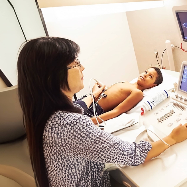
As with all surgery patients, Arden underwent routine imaging that produced two-dimensional pictures of her kidney, which showed the tumor was sitting dangerously close to the organ’s main blood supply. This presented Kolon with a problem: What was the best way to access the tumor?
Luckily, there was a new development at CHOP: the creation of the Children’s Hospital Additive Manufacturing for Pediatrics (CHAMP) Lab in the Department of Radiology. Run by Elizabeth Silvestro, MSE, and Raymond Sze, MD, MAMS, the CHAMP Lab focuses on printing 3D models. Silvestro and Sze knew that using 3D models could open the door to finding innovative ways to treat patients, and they actively solicited ideas from a wide range of clinicians across the hospital.
After viewing a presentation that included 3D printed models used in urology research, Kolon saw an opportunity, and with Arden in mind, he approached Susan Back, MD, pediatric radiologist, with a question: “Can we use this to take a better look at tumors and the surrounding tissue?”
Printing 3D replicas of organs
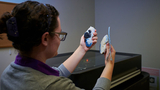
Silvestro and Back used Arden’s 2D imaging to create a 3D model of her kidney: “We took the images,” explains Back, “segmented the anatomy and used the lab’s advanced programming to print it.” The kidney model could be opened up to directly visualize the tumor and its relationship to the arteries, veins and other structures, allowing the anatomy to be studied from a new perspective.
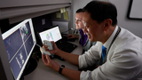
“We wanted to save as much healthy kidney tissue as possible to avoid her going on dialysis,” says Kolon. “This 3D model allowed me to see the critical blood supply to the kidney and to the tumor. It allowed me to have access to views I didn’t have before. I carried the model around and looked at it from different viewpoints and looked at the blood supply. This made a tremendous difference.” In April 2018, Kolon performed surgery that removed the tumor and spared the rest of Arden’s kidney.
Arden’s recovery from the six-hour surgery was difficult, but she’s now doing great. “She loves to dance and do gymnastics,” says Tray. Without the team’s innovative approach that resulted in success at the surgery, “Arden would not have been able to do everything she’s doing now.”
‘This is possible for so many patients’
If Arden was anywhere other than CHOP, most likely a partial kidney removal wouldn’t even have been attempted. Since her surgery, the 3D printing technology has been used for many other patients, such as a boy who, like Arden, developed a tumor in the middle of his one remaining kidney and came to CHOP for a second opinion. “It was thought to be non-operable,” says Kolon, “but we were able to remove the entire tumor and reconstruct the kidney.”
“The collaboration between the clinician, radiologist and engineer has been an important part of creating these models,” says Back. “Understanding how a surgeon uses the model allows us to plan and optimize the imaging study in order to create a presurgical print.”
3D modeling changes the equation for other organs, too. Doctors at another hospital were going to remove a young girl’s bladder because of cancerous tumors. The family came to CHOP for a second opinion. “We made a 3D model,” says Kolon, “and I was able to do the procedure through a scope, and she is tumor-free with her bladder saved. The success we’ve had is allowing us to have the confidence to say organ-sparing surgery is possible for so many patients.”
As an added benefit, when a surgeon has the opportunity to closely examine a 3D model and make a plan beforehand, there’s a good chance the operation will go faster, meaning the patient will spend less time under anesthesia.
More uses of 3D printing in medicine
Using patient-specific 3D models to help surgeons prepare for complex procedures is a striking use of 3D printing technology, but CHOP’s CHAMP Lab knows that is just scratching the surface.
For one, 3D models can help trainees learn skills. For example, for an operation that requires cutting and reconstructing the pelvic bone, Silvestro can craft a 3D model using materials that simulate bone, so a surgeon-in-training can get a sense of what it will actually feel like to do the procedure. Such knowledge of and experimentation with materials is where Silvestro’s expertise shines through.
3D models are also used to prototype new pediatric-specific biomedical devices. The CHAMP Lab developed a device that allows clinicians in the Division of Plastic and Reconstructive Surgery to better measure the nasal cavity of children with a cleft lip or palate, and there are many other devices in the testing phase.
CHAMP Lab team members are thrilled by what they’ve been able to accomplish to date, and they look forward to the next innovative idea for using 3D printing technology to enhance patient care. The team sees endless possibilities. As Sze notes with excitement, “We’re getting requests from across the hospital!”
Featured in this article
Specialties & Programs
As her 5-year-old daughter Arden prepared to face the second surgery of her young life, Tray Sullivan MacPhee’s anxiety grew. “I wasn’t yet Googling ‘kids on dialysis,’” Tray says. But she feared that would be Arden’s future.
That’s because back when she was 18 months old, Arden was diagnosed with a cancer of the kidneys called Wilms tumor, a disease that occurs more often in children than adults. Before she turned 2, Arden underwent 12 rounds of chemotherapy at Children’s Hospital of Philadelphia (CHOP). After chemo, Thomas Kolon, MD, pediatric urologist, performed surgery to remove the kidney that contained a large cancerous mass, invasive into her central blood supply and drainage system.
For three years, everything was fine — until cancer was detected in Arden’s other kidney. “It was more shocking than the first time,” says Tray.
If Arden’s remaining kidney had to be removed, she would require dialysis to clean her blood. Not only does each dialysis treatment take several hours, it needs to happen every few days indefinitely until a possible kidney transplant — a challenging treatment for anyone, let alone a young child. Both her family and medical team hoped for a way to remove the cancer while saving the kidney.
As with all surgery patients, Arden underwent routine imaging that produced two-dimensional pictures of her kidney, which showed the tumor was sitting dangerously close to the organ’s main blood supply. This presented Kolon with a problem: What was the best way to access the tumor?
Luckily, there was a new development at CHOP: the creation of the Children’s Hospital Additive Manufacturing for Pediatrics (CHAMP) Lab in the Department of Radiology. Run by Elizabeth Silvestro, MSE, and Raymond Sze, MD, MAMS, the CHAMP Lab focuses on printing 3D models. Silvestro and Sze knew that using 3D models could open the door to finding innovative ways to treat patients, and they actively solicited ideas from a wide range of clinicians across the hospital.
After viewing a presentation that included 3D printed models used in urology research, Kolon saw an opportunity, and with Arden in mind, he approached Susan Back, MD, pediatric radiologist, with a question: “Can we use this to take a better look at tumors and the surrounding tissue?”
Printing 3D replicas of organs
Silvestro and Back used Arden’s 2D imaging to create a 3D model of her kidney: “We took the images,” explains Back, “segmented the anatomy and used the lab’s advanced programming to print it.” The kidney model could be opened up to directly visualize the tumor and its relationship to the arteries, veins and other structures, allowing the anatomy to be studied from a new perspective.
“We wanted to save as much healthy kidney tissue as possible to avoid her going on dialysis,” says Kolon. “This 3D model allowed me to see the critical blood supply to the kidney and to the tumor. It allowed me to have access to views I didn’t have before. I carried the model around and looked at it from different viewpoints and looked at the blood supply. This made a tremendous difference.” In April 2018, Kolon performed surgery that removed the tumor and spared the rest of Arden’s kidney.
Arden’s recovery from the six-hour surgery was difficult, but she’s now doing great. “She loves to dance and do gymnastics,” says Tray. Without the team’s innovative approach that resulted in success at the surgery, “Arden would not have been able to do everything she’s doing now.”
‘This is possible for so many patients’
If Arden was anywhere other than CHOP, most likely a partial kidney removal wouldn’t even have been attempted. Since her surgery, the 3D printing technology has been used for many other patients, such as a boy who, like Arden, developed a tumor in the middle of his one remaining kidney and came to CHOP for a second opinion. “It was thought to be non-operable,” says Kolon, “but we were able to remove the entire tumor and reconstruct the kidney.”
“The collaboration between the clinician, radiologist and engineer has been an important part of creating these models,” says Back. “Understanding how a surgeon uses the model allows us to plan and optimize the imaging study in order to create a presurgical print.”
3D modeling changes the equation for other organs, too. Doctors at another hospital were going to remove a young girl’s bladder because of cancerous tumors. The family came to CHOP for a second opinion. “We made a 3D model,” says Kolon, “and I was able to do the procedure through a scope, and she is tumor-free with her bladder saved. The success we’ve had is allowing us to have the confidence to say organ-sparing surgery is possible for so many patients.”
As an added benefit, when a surgeon has the opportunity to closely examine a 3D model and make a plan beforehand, there’s a good chance the operation will go faster, meaning the patient will spend less time under anesthesia.
More uses of 3D printing in medicine
Using patient-specific 3D models to help surgeons prepare for complex procedures is a striking use of 3D printing technology, but CHOP’s CHAMP Lab knows that is just scratching the surface.
For one, 3D models can help trainees learn skills. For example, for an operation that requires cutting and reconstructing the pelvic bone, Silvestro can craft a 3D model using materials that simulate bone, so a surgeon-in-training can get a sense of what it will actually feel like to do the procedure. Such knowledge of and experimentation with materials is where Silvestro’s expertise shines through.
3D models are also used to prototype new pediatric-specific biomedical devices. The CHAMP Lab developed a device that allows clinicians in the Division of Plastic and Reconstructive Surgery to better measure the nasal cavity of children with a cleft lip or palate, and there are many other devices in the testing phase.
CHAMP Lab team members are thrilled by what they’ve been able to accomplish to date, and they look forward to the next innovative idea for using 3D printing technology to enhance patient care. The team sees endless possibilities. As Sze notes with excitement, “We’re getting requests from across the hospital!”
Recommended reading
Radiology
The Department of Radiology at CHOP provides safe, specialized imaging services for infants, children and pregnant women.
Contact us
Division of Urology