Tetralogy of Fallot (TOF): Mason's Story
Tetralogy of Fallot (TOF): Mason's Story
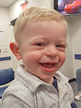
Mason is a happy toddler who delights in blowing kisses. His parents think many of those should go to his medical team at Children’s Hospital of Philadelphia (CHOP), where his heart was repaired as a baby.

Kelly and Matt went to their 20-week ultrasound as expectant parents, excited to learn the sex of their baby. The technician told them they were having a boy, but also noticed there might be something wrong with his heart. The image wasn’t clear enough to be sure, so their doctor referred them to Children’s Hospital of Philadelphia for a higher-resolution ultrasound and an echocardiogram.
Diagnosis with congenital heart disease (CHD)
At CHOP, Kelly and Matt learned that their baby had a combination of anatomical problems in his heart, which together are known as tetralogy of Fallot (TOF). He had what appeared to be a large hole between the two bottom chambers of his heart, a problem known as ventricular septal defect (VSD), which allowed oxygenated and deoxygenated blood to mix. His aorta was positioned over this defect (overriding aorta), instead of over the left ventricle. And his pulmonary valve and pulmonary artery were constricted, partially blocking the flow of blood from his heart to his lungs.
He would need surgery soon after birth to repair his heart, and Kelly would need to deliver at CHOP to ensure the new baby had access to immediate care. They were stunned. “I was in a complete daze,” Matt remembers.
More About TOF
For the remainder of her pregnancy, Kelly went to CHOP for monthly ultrasounds to monitor her baby’s heart. She made arrangements to deliver at CHOP’s Garbose Family Special Delivery Unit, the first birth facility in a pediatric hospital designed for healthy mothers carrying babies with known birth defects. Moms can stay close, and babies are treated immediately without having to be transported.
Mason was born in early December. Kelly’s delivery team was in the room with her, and in an adjoining room, separated by a window, Mason’s medical team stood ready.
Mason came out breathing, his condition relatively stable. The delivery team briefly laid him on Kelly’s chest, then passed him through the window to his own medical team. They cleaned him up, put catheters to give him medications, then brought him back in so that his parents could hold him before he was taken to the Cardiac Intensive Care Unit (CICU).
Heart surgery at 5 days old
On the following Tuesday, when he was 5 days old, Mason had surgery to repair his heart. Thomas Spray, MD, Chief of the Division of Cardiothoracic Surgery, accomplished three objectives in the procedure. He reduced the size of the hole between the two ventricles, inserting a patch to plug the opening. He placed a transannular patch in the pulmonary valve to open it more fully, and he enlarged the pulmonary artery. He did all of this on a tiny infant heart.
After the surgery, Kelly and Matt met with Meryl Cohen, MD, who would lead Mason’s care team moving forward. She has been his cardiologist ever since, and both parents think the world of her. Two weeks after his operation, Mason was well enough to go home.
New problems
At a follow-up visit a few weeks later, a scan picked up a muscle bundle growing in Mason’s right ventricle. By its thickness and position, it was blocking outflow from his heart.
When Mason was 4 months old, Jonathan Rome, MD, director of CHOP’s Cardiac Catheterization Laboratory, performed a diagnostic cardiac catheterization. Mason’s cardiac team, including Drs. Cohen, Spray and Rome, decided open heart surgery was required to remove the muscle bundle. In the same procedure, Dr. Spray would make an additional repair to the VSD to completely close the hole.
The operation was a success, but a chest X-ray in the days following the procedure showed that blood flow to the left lung was reduced. The left pulmonary artery had gotten smaller or had become twisted.
Once Mason had recovered from the surgery, Dr. Rome used catheterization to insert a stent in the artery to widen it and allow more blood to flow through it. He selected an adult stent, modified to fit a baby’s artery, so that it could be widened every few years as Mason grew. A stent can be widened using catheterization until it reaches its maximum size. If the artery needs a larger stent after that, as a child grows into adulthood, open heart surgery is required to replace it. By using a modified version of an adult stent, Dr. Rome was trying to prolong the need for a third open heart surgery when Mason was older.
A happy toddler

That catheterization was the last procedure Mason needed as a baby. He went home to a normal life. His parents bring him back to CHOP every six months to be examined by Dr. Cohen. He generally has an echocardiogram and an electrocardiogram (EKG) as part of those visits.
Now a toddler, Mason is talking up a storm, running and climbing on anything and everything. He loves being in the water, playing outside, patting the family’s dog and blowing kisses. He’s a happy little boy who usually has a big smile on his face. Matt says:
“CHOP is amazing. Mason's doctors, the nurses, the assistants – they are all amazing. There’s not a day that goes by that I don’t think of them.”