Division of Ophthalmology

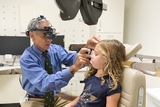
If your child has any of a wide range of eye and vision problems — both common and rare — they will find expert care from pediatric eye specialists in the Division of Ophthalmology at Children's Hospital of Philadelphia (CHOP).
We provide comprehensive evaluation, coordinated care and cutting-edge treatment. As one of the leading pediatric ophthalmology centers in the United States, children travel to CHOP for treatment of eye misalignment, abnormal eye movements, lazy eye, poor visual function and less common pediatric eye conditions such as glaucoma, cataracts, tearing problems or droopy eyelids.
We also offer special testing not routinely available for children elsewhere.
Adult patients with strabismus, double vision and other eye movement problems are also treated by CHOP ophthalmologists.
How we serve you
In addition to the specialty clinics listed below, we also offer an Optometry Clinic, a Neuro-ophthalmology Clinic, an Oculoplastics and Tearing Disorders Clinic, and a Pediatric Retina Clinic.
Conditions we treat
Learn more about some of the many ocular conditions we treat:
-
Amblyopia (lazy eye) -
Blepharitis -
Blocked tear duct (dacryostenosis) -
Cataracts in children -
Chalazion -
Childhood glaucoma -
Conjunctivitis (pink eye) -
Corneal abrasions -
Crossed-eyes (strabismus) -
Functional vision -
Keratitis -
Myasthenia gravis -
Neurofibromatosis type 1 -
Neurofibromatosis type 2 -
Optic neuritis -
Pseudotumor cerebri syndrome (PTCS) -
Refractive errors in children -
Retinopathy of prematurity -
Stye (hordeolum) -
Syndromic craniosynostosis -
Thyroid eye disease -
Uveitis

Why choose CHOP for eye and vision care
Our team provides safe, high-quality and effective ophthalmic care at nine convenient locations. We use the latest diagnostic testing and ophthalmic imaging, and pursue clinical research to improve outcomes for all children with eye conditions.

Meet your team
Our team of ophthalmologists, surgeons, optometrists, orthoptists, nurse practitioners and certified ophthalmic technicians is dedicated to providing the best possible care for your child's eye or vision condition.

Our locations
Our pediatric eye specialists provide care at several CHOP Care Network locations throughout the region. We are pleased to offer late afternoon and Saturday hours at many of our locations, including the Buerger Center for Advanced Pediatric Care, Virtua Voorhees, and our King of Prussia, Exton and Brandywine Valley specialty care centers.

Our research
Our doctors and researchers are actively involved in studies on retinopathy of prematurity, strabismus, cataracts, eye growth, myopia, amblyopia and many other related areas. Our physicians have published widely on topics related to pediatric ophthalmology.

Eye care resources
We have collected helpful resources on pediatric eye care so you can feel confident in the care you're providing your child.

What to expect at your child's appointment
See what you need to bring with you and what your child can expect at your appointment, including possible same-day testing.

Your child's eye surgery
We answer your questions on scheduling, referrals and how to prepare your child for their surgery.
Your donation changes lives
A gift of any size helps us make lifesaving breakthroughs for children everywhere.


