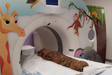

For the first time in the hospital’s 163-year history, Children’s Hospital of Philadelphia (CHOP) took on a 2,000-year-old case. Radiologists conducted a computerized tomography, or CT, scan on a mummified human, provided by Penn Museum. The mummy is assumed to be a girl based on the decoration of her elaborately painted plaster and linen shroud. She lived around 270–280 CE during the Roman Empire in Egypt.
Previous X-rays done on the mummy showed anatomical inconsistencies, which made it difficult to determine the child’s age or identify a cause of death. Penn Museum and CHOP believe the CT images will shed light on how the girl lived and why she died at a young age.
Featured in this article
Specialties & Programs

For the first time in the hospital’s 163-year history, Children’s Hospital of Philadelphia (CHOP) took on a 2,000-year-old case. Radiologists conducted a computerized tomography, or CT, scan on a mummified human, provided by Penn Museum. The mummy is assumed to be a girl based on the decoration of her elaborately painted plaster and linen shroud. She lived around 270–280 CE during the Roman Empire in Egypt.
Previous X-rays done on the mummy showed anatomical inconsistencies, which made it difficult to determine the child’s age or identify a cause of death. Penn Museum and CHOP believe the CT images will shed light on how the girl lived and why she died at a young age.
Contact us
Department of Radiology