
Talipes (known as clubfoot) is one of the most common congenital foot and ankle problems, occurring in about one in 1,000 live births and in boys twice as often than in girls. The past decade has seen increased prenatal detection of clubfoot, thanks to imaging advances and increases in sonographic screening during pregnancy.
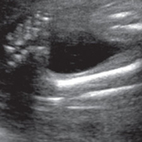
Diagnosis typically occurs via ultrasound at 20 weeks gestation and is based on visualization of the foot in a plantarflexed and internally rotated position so the sole faces inward. The Achilles tendon is contracted and the calcaneus is proximally displaced from its normal position, there may be a crease on the bottom of the foot, and the affected foot, calf and hallux may be smaller than the unaffected side. Roughly half of all cases are bilateral.
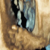
Our world-leading fetal diagnosis and imaging program ensures the most accurate diagnosis and facilitates early consultation between families and our orthopaedic team. Families undergo a comprehensive evaluation that includes a 3-D fetal ultrasound and sometimes an ultrafast fetal MRI, depending on the diagnosis. Following the diagnostic imaging, our maternal-fetal medicine specialists and orthopaedic surgeons meet with families to discuss findings and their implications, postnatal management, treatment options, and long-term prognosis.
Establishing this connection and relationship before birth is enormously helpful to both families and our clinical team. Families leave knowing what to expect once the baby is born, and have a number to call if any questions come up.
Treatment and follow-up care
Because the severity of the condition and the best course of treatment are difficult to predict until the child is born, our orthopaedic team makes plans to see families within the first few weeks of birth to begin treatment.
Our treatment approach follows the Ponseti method. Serial casting is the first step and involves gentle manipulation of the child’s foot and application of a long leg cast. Casts are changed weekly. Typically five to seven casts are needed for correction. In most cases, the Achilles tendon will need to be cut (Achilles tenotomy) in order to complete the correction. This is typically performed in the operating room under local anesthesia. The child is casted for another three to four weeks and then fitted for a brace that will be worn 23 hours a day for three months and then at night until age 4. Our goal is to slightly overcorrect the foot so that it will look normal at the end of treatment.
Regardless of treatment, a clubfoot is usually shorter and wider than an unaffected foot, the calf is usually smaller, and the affected leg may be slightly shorter than a leg without a clubfoot. But with early care from a multidisciplinary team, outcomes are usually excellent. The foot is usually corrected before the child starts crawling and walking. Most children have mobile, painless and flexible feet, can wear normal shoes, and are able to lead a healthy and active life and participate in sports. Our team follows children throughout childhood, until they are stable and there is no risk of recurrence.
Clubfoot and spina bifida
While clubfoot is typically an isolated anomaly, other conditions (such as spina bifida or arthrogryposis) may be associated. Our Center for Fetal Diagnosis & Treatment is highly skilled in the prenatal diagnosis of these and other diagnoses that may be associated with clubfoot. The center is a pioneer in fetal surgery for spina bifida, a treatment that can minimize the incidence and severity of clubfoot and improve the chances that a child will be able to walk independently.
Whether closure of the spinal defect occurs before or after birth, children who have clubfoot from spina bifida tend to have more recurrence of clubfoot and need more extensive treatment than those without spina bifida. These children can also have complex medical issues involving multiple body systems such as the central nervous system, musculoskeletal system, and urinary and intestinal systems. Our multidisciplinary Spina Bifida Program provides coordinated care and long-term management for infants, children and teens to ensure the best outcomes.
Featured in this article
Specialties & Programs
Talipes (known as clubfoot) is one of the most common congenital foot and ankle problems, occurring in about one in 1,000 live births and in boys twice as often than in girls. The past decade has seen increased prenatal detection of clubfoot, thanks to imaging advances and increases in sonographic screening during pregnancy.
Diagnosis typically occurs via ultrasound at 20 weeks gestation and is based on visualization of the foot in a plantarflexed and internally rotated position so the sole faces inward. The Achilles tendon is contracted and the calcaneus is proximally displaced from its normal position, there may be a crease on the bottom of the foot, and the affected foot, calf and hallux may be smaller than the unaffected side. Roughly half of all cases are bilateral.
Our world-leading fetal diagnosis and imaging program ensures the most accurate diagnosis and facilitates early consultation between families and our orthopaedic team. Families undergo a comprehensive evaluation that includes a 3-D fetal ultrasound and sometimes an ultrafast fetal MRI, depending on the diagnosis. Following the diagnostic imaging, our maternal-fetal medicine specialists and orthopaedic surgeons meet with families to discuss findings and their implications, postnatal management, treatment options, and long-term prognosis.
Establishing this connection and relationship before birth is enormously helpful to both families and our clinical team. Families leave knowing what to expect once the baby is born, and have a number to call if any questions come up.
Treatment and follow-up care
Because the severity of the condition and the best course of treatment are difficult to predict until the child is born, our orthopaedic team makes plans to see families within the first few weeks of birth to begin treatment.
Our treatment approach follows the Ponseti method. Serial casting is the first step and involves gentle manipulation of the child’s foot and application of a long leg cast. Casts are changed weekly. Typically five to seven casts are needed for correction. In most cases, the Achilles tendon will need to be cut (Achilles tenotomy) in order to complete the correction. This is typically performed in the operating room under local anesthesia. The child is casted for another three to four weeks and then fitted for a brace that will be worn 23 hours a day for three months and then at night until age 4. Our goal is to slightly overcorrect the foot so that it will look normal at the end of treatment.
Regardless of treatment, a clubfoot is usually shorter and wider than an unaffected foot, the calf is usually smaller, and the affected leg may be slightly shorter than a leg without a clubfoot. But with early care from a multidisciplinary team, outcomes are usually excellent. The foot is usually corrected before the child starts crawling and walking. Most children have mobile, painless and flexible feet, can wear normal shoes, and are able to lead a healthy and active life and participate in sports. Our team follows children throughout childhood, until they are stable and there is no risk of recurrence.
Clubfoot and spina bifida
While clubfoot is typically an isolated anomaly, other conditions (such as spina bifida or arthrogryposis) may be associated. Our Center for Fetal Diagnosis & Treatment is highly skilled in the prenatal diagnosis of these and other diagnoses that may be associated with clubfoot. The center is a pioneer in fetal surgery for spina bifida, a treatment that can minimize the incidence and severity of clubfoot and improve the chances that a child will be able to walk independently.
Whether closure of the spinal defect occurs before or after birth, children who have clubfoot from spina bifida tend to have more recurrence of clubfoot and need more extensive treatment than those without spina bifida. These children can also have complex medical issues involving multiple body systems such as the central nervous system, musculoskeletal system, and urinary and intestinal systems. Our multidisciplinary Spina Bifida Program provides coordinated care and long-term management for infants, children and teens to ensure the best outcomes.
Contact us
Richard D. Wood Jr. Center for Fetal Diagnosis and Treatment