Sophia’s Story: Fetal Surgery to Remove a Sacrococcygeal Teratoma
Sophia’s Story: Fetal Surgery to Remove a Sacrococcygeal Teratoma
In April 2020, as the COVID-19 pandemic upended lives, the economy and society, Kristen and Shawn were focused on their 20-week ultrasound, where they would learn more about their first child, particularly if the baby was a girl or a boy. They never could’ve imagined what was about to unfold. From that ultrasound appointment to a successful fetal surgery to welcoming their baby girl, Sophia, read on to follow their journey.

Shawn wasn’t allowed to be present for his first child’s ultrasound due to COVID, so he waited in his car in the parking lot, eager to learn whether the baby was a girl or a boy. But when the OB-GYN asked his wife, Kristen, to have him come in to discuss the ultrasound results, it was clear something was wrong. The OB said he saw something in the child’s pelvic area, but he wasn’t sure what it was. He referred them to a high-risk specialist for maternal-fetal medicine.
The couple was terrified in the week between the appointments. When they met with the maternal-fetal medicine (MFM) specialist in Voorhees, N.J., not far from their home in Williamstown, N.J., she confirmed the baby had a sacrococcygeal teratoma (SCT), a potentially life-threatening tumor located at the base of the tailbone.
SCTs can grow rapidly, so it is critical to go to a fetal treatment center with extensive experience. The MFM referred the couple to the Richard D. Wood Jr. Center for Fetal Diagnosis and Treatment (CFDT) at Children’s Hospital of Philadelphia, which has one of the greatest experiences in the world with fetal management of SCT.
A clear SCT diagnosis and a plan of action
From their first appointment at CHOP, Kristen says she was very impressed. What struck her most was the nurse coordinator who guided them through the day-long evaluation.
“She organized the entire day for us,” says Kristen, a special education teacher. “It was a stressful day, with a lot of tests, but having such a clear plan was very helpful in terms of my nerves.”
Kristen underwent a thorough, day-long evaluation at the Center to confirm the diagnosis. Tests included fetal ultrasound, ultra-fast fetal MRI, and fetal echocardiogram. At the end of the day, Kristen and Shawn met with the center’s team to discuss the details of the tumor. It was a large, type 2 SCT, which meant there was a small portion of the tumor growing inside the body and a larger portion growing outside of the body.
Large teratomas that grow into the abdomen can compress adjacent developing organs, the airway in the neck or intra-abdominal structures. They can also have consequences in terms of their blood supply. If they're solid and richly supplied with blood, the teratoma can steal blood away from the developing fetus and placenta and cause cardiovascular failure.
If the tumor kept growing at its current pace, the fetus would go into heart failure. The Center collaborates with CHOP’s specialized Fetal Heart Program to follow SCTs extremely closely through ongoing fetal echocardiograms. This allows the team to monitor how hard the fetus’s heart is working and whether the fetus is going into heart failure (also called fetal hydrops). If it looks like the fetus is going into heart failure, the team can either deliver the baby at an earlier gestational age and treat the baby outside the womb, or perform fetal surgery to remove part of the tumor and reduce the stress on the fetal heart.
“We were really scared, and we didn’t know what the outcome would be,” says Kristen. “We just took it day by day, week by week. But I had a gut feeling that I was going to need fetal surgery.”
Kristen’s instinct was right. At 24 weeks, the tumor was still growing. The fetal combined cardiac output — a measurement of how hard the heart is working — was very high, and the tumor was pressing on other organs like the kidneys. Concerned that Kristen would develop maternal mirror syndrome — in which a mother’s health mirror’s that of her sick fetus — and that the fetus would not survive, the team offered to perform fetal surgery.
Successful fetal surgery for sacrococcygeal teratoma
The day of fetal surgery also happened to be Kristen’s birthday. The care team went above and beyond to make her feel special despite all she was going through. They made her a playlist of her favorite songs to listen to in the operating room and sang her happy birthday before she went under anesthesia.
“Obviously they are trained to help medically, but they also helped emotionally,” says Kristen of the fetal team. “They made me feel so relaxed. It wasn’t as scary as I thought because these people calmed me down so much.”
Fetal surgeon William H. Peranteau, MD, and the Center’s large, highly specialized team of pediatric/fetal surgeons, maternal fetal medicine specialists, fetal surgery operating room nurses, and obstetric and fetal anesthesiologists, safely removed about 90% of the tumor and closed the uterus in the hopes of prolonging the pregnancy as long as possible so the fetus could continue to grow. Throughout the procedure, a dedicated fetal cardiologist performed continuous fetal echocardiogram monitoring.
Later that day, a repeat echocardiogram showed that fetal cardiac output had already improved. Kristen returned to CHOP twice a week for follow-up appointments, which showed continued improvement in the fetus’s heart function.
SDU birth, N/IICU care and discharge home
Fetal surgery gave Kristen’s daughter, Sophia 11 more weeks in the womb, improving her heart function and her chances of survival. Around 34 weeks’ gestation, an ultrasound revealed that the tumor was growing inside and outside the fetus’s body again, so, on July 20, 2020, Kristen underwent a planned C-section in CHOP’s Garbose Family Special Delivery Unit (SDU). The SDU was created specifically for families like hers, whose children require surgery and/or intensive care immediately after birth.

Delivering at CHOP
Learn about what sets CHOP’s Garbose Family Special Delivery Unit apart in the care of mothers carrying babies with prenatally diagnosed birth defects.
Two weeks after the birth, Dr. Peranteau surgically removed the rest of the tumor and the tailbone — which helps prevent tumor recurrence — and reconstructed Sophia’s bottom. While Sophia stayed in the Harriet and Ronald Lassin Newborn/Infant Intensive Care Unit (N/IICU) for about a month recovering, the N/IICU team supported the family in myriad ways.
When they had to take breaks from being in the N/IICU, a camera at Sophia’s bedside enabled them to view her in real-time through their smartphones, giving them peace of mind. They gave her grandparents access to the camera, so they could see Sophia, as well. A child life specialist worked one-on-one with the parents to create a milestone bracelet for Sophia; for each struggle she overcame — e.g., getting her IV — a bead was added to the bracelet.
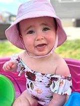
Today, Sophia is doing well. She was discharged from the hospital weighing about 7 lbs., and now, 9 months old, is growing well. The family returns to CHOP for follow-up appointments with surgery, oncology, neurology and urology. Knowing she is being followed by an expert team of specialists is a huge relief to Sophia’s parents.