What is hemifacial microsomia?
Hemifacial microsomia is a congenital condition in which the tissues on one side of the face are underdeveloped. It primarily affects the ear, mouth and jaw areas, though it may also involve the eye, cheek, neck and other parts of the skull, as well as nerves and soft tissue. In 10–15% of cases, both sides of the face are affected, often asymmetrically.
Hemifacial microsomia is the second most common facial birth defect behind cleft lip and palate, affecting one in every 3,500 to 4,000 births. The areas of the face that are affected at birth will typically remain similarly proportionally asymmetric throughout growth and development, neither worsening nor getting better.
Hemifacial microsomia has several other names, including craniofacial microsomia, Goldenhar syndrome, first and second branchial arch anomaly, branchial arch syndrome, facioauriculovertebral syndrome, oculoauriculovertebral spectrum, and lateral facial dysplasia.
Goldenhar Syndrome is a variant of hemifacial microsomia in which there are spine anomalies and/or small benign tumors of the eye or eyelid called dermoids or lipodermoids.
Watch this short video to learn more about hemifacial microsomia.
Causes of hemifacial microsomia
It is unclear what causes hemifacial microsomia. Recent studies have identified several genes associated with this condition, such as GATA3, SH3B2 and FOXI3. Historically, there were theories that the malformation process starts in the first trimester of pregnancy and may be caused by a vascular problem leading to poor blood supply to the fetus’ face during early development, but this idea has no experimental support. We do not believe that the facial anomalies are triggered by a mother’s actions or diet.
In most cases, the condition is not inherited and happens by chance. In a small minority of cases, a child may inherit the condition from their parents. An adult with hemifacial microsomia has about a 3% chance or less of having a child with the same condition.
Signs and symptoms of hemifacial microsomia
Symptoms of hemifacial microsomia range from severe to barely noticeable and depend greatly on the degree of deformity and how much of the face is involved. A child with a mild form of hemifacial microsomia may have a slightly smaller jaw and a skin tag in front of a normal-looking ear. In more severe forms, a child’s face may appear much smaller on one side, with an abnormally shaped or absent ear.
One of the ways clinicians describe and assess the severity of hemifacial microsomia is the OMENS classification. The OMENS classification examines the function and appearance of each of the following, looking for characteristics commonly associated with hemifacial microsomia:
- Orbit (eye socket): small and underdeveloped eyes with impaired vision; absent or unformed eye; growths on the eye; one eye appearing smaller than the other
- Mandible (the jaw bones): underdeveloped upper and lower jaw on one side; crooked jaw; missing, misaligned or overcrowded teeth; cleft lip and/or cleft palate; limited opening or closing of the mouth
- Ear: small skin tags; misshapen or missing external ear; absent or abnormal development of the ear canal resulting in partial or total hearing loss
- Nerves: ranging from mild weakness to partial or full facial paralysis
- Soft tissues (skin, muscle, fat, tendons and ligaments): asymmetry of facial volume and movement, due to absence of or smaller muscle mass; absent fatty tissue
Most children with hemifacial microsomia have facial anomalies but no other major medical issues. Less commonly, babies born with hemifacial microsomia may also have other health problems such as malformed vertebrae, heart defects or abnormally shaped kidneys.
Testing and diagnosis
The diagnosis of hemifacial microsomia can be made before or after birth. Some of the abnormal facial features are visible during prenatal ultrasound. If you are pregnant and your unborn child has these distinctive characteristics, you may be referred to CHOP’s Center for Fetal Diagnosis and Treatment.
Most children are not diagnosed with hemifacial microsomia until after birth. In this case, experts from CHOP’s Craniofacial Program will evaluate your child. Experienced physicians will make the diagnosis based on your child’s appearance: the mandibular (jaw) deformity is the hallmark of hemifacial microsomia and is classified based on the development of the jaw. In the mildest formation, the mandible is nearly normal and only slightly hypoplastic (underdeveloped), while in the most severe cases, a portion of the jaw is missing on the affected side.
Some of the facial characteristics of hemifacial microsomia mimic those seen in children with Treacher Collins syndrome, but hemifacial microsomia differences are typically one-sided or asymmetric, whereas in Treacher Collins, characteristics are similar on both sides of the face.
Diagnostic tests such as X-rays and CT scans may also be used to better examine your child’s bone and cartilage structure in order to make appropriate treatment recommendations.
Surgical treatment options for hemifacial microsomia
The treatment of hemifacial microsomia varies from patient to patient, depending on the severity of the condition and long-term needs of the child. Consultation with an experienced craniofacial team is extremely important in achieving the best outcomes for your child.
At CHOP — where we have expertise in every pediatric specialty — your child will have access to a multidisciplinary healthcare team and coordinated care through our Craniofacial Program. We will work with you to prioritize your child’s needs and establish a comprehensive treatment plan that addresses both physical and psychosocial needs, including planning for staged surgical repair and reconstruction.
Timing can be a critical factor in treatment of hemifacial microsomia. In some cases, treatment can wait until your child reaches certain developmental milestones.
At CHOP we also bring the latest research to the care of hemifacial microsomia. We have carried out decades-long clinical studies of how to restore facial and jaw symmetry, including updating the OMENS classification to better describe the condition. Our Advancing Craniofacial Treatment with Genomics and Gene Therapy Frontier Program specifically seeks to address the gap in knowledge in genetics of hemifacial microsomia and aims to better describe the natural history of this condition paired with better genomic diagnosis in the future.
Supportive treatment at birth
If your child is born with hemifacial microsomia, they may require respiratory support or a tracheostomy if the jaw is severely deficient. In most cases, your child’s airway can be managed conservatively.
Due to the presence of the jaw deformity and clefts, your child may experience feeding difficulties. Supplemental feedings through a nasogastric tube may be needed to support growth and weight gain.
If facial paralysis or eyelid abnormalities are present, eye closure may be incomplete and eye protection must be provided either via lubricants or surgical procedures.
Surgical treatment
As your child with hemifacial microsomia grows, surgical treatment may be needed based on the severity and area affected. Not all children with hemifacial microsomia have problems in all of these areas.
Below are some of the interventions your child may need. If you have any questions about treatment for your child, please talk to your child’s healthcare team or call the Division of Plastic, Reconstructive and Oral Surgery at 215-590-2208.
Ears: Some children with abnormally shaped or missing ears may choose to have a series of reconstructive surgeries to make the ear appear more normal. The first surgery typically occurs after age 6, when your child’s ears have almost reached adult size. Another option is to make an artificial or prosthetic ear, which also requires several surgeries. Learn more about how ear deformities are treated by the Division of Plastic, Reconstructive and Oral Surgery.
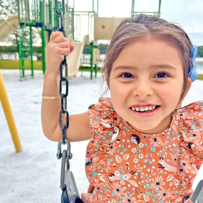
Watch the inspiring story of a little girl with hemifacial microsomia who had ear reconstruction surgery at CHOP.
Transcript Transcript - Eyelid differences: For children with eyelid differences, surgical procedures to repair or reposition the upper and lower eyelid may be required.
- Soft tissue deficiencies: Children with skin, cheek and other soft tissue deficiencies may need augmentation procedures such as fat grafting or tissue transfer.
- Lateral facial cleft/cleft lip and palate: Babies born with cleft lip or palate can have surgical repairs done during the child’s first year. Cleft lip repair is typically performed when your child is 3 to 6 months old, while cleft palate surgery is generally performed when your child is about a year old. A lateral facial cleft is one of the most severe deficiencies found with hemifacial microsomia. It requires a staged reconstruction, similar to the process used in routine repair of cleft lip and palate. In this procedure, surgeons will create a ring of muscle around your child’s mouth, connecting the corners and drawing up the lateral line of the lower lip. This reconstructive procedure also helps with feeding and speech.
- Bony deficiencies: In mild cases of bony deficiencies, no treatment may be needed. In more severe cases, surgery may be required. Two of the most commonly performed procedures include distraction lengthening of the mandible (most common) and reconstruction of the mandible with a rib or free vascularized fibula graft (less common).
For a distraction lengthening of the mandible, a surgeon cuts the mandible (jaw) in the center of its deficient region and implants a small device that allows the two bone segments to be distracted (pulled apart), creating a gap in the bone. New bone begins to form in the gap of the small jaw and the device is slowly opened until the the jaw reaches the appropriate size. When the jawbone has been adjusted, it improves facial form and the way the top and bottom teeth fit together. Mandibular distraction may need to be repeated as your child grows.
As is the case for any patient with a complex craniofacial deformity, individual treatment varies depending upon the degree of involvement of the various structures. Your child’s individual treatment plan may vary from others similarly affected due to a variety of other factors. It is important that you see an experienced craniofacial team to manage and assess your child’s condition.
Follow-up care
As your child with hemifacial microsomia grows into adolescence, they should continue to be monitored by experienced physicians who can adjust treatment plans as needed.
Because multiple body systems may be involved in hemifacial microsomia, continued monitoring for complications and any treatment as needed are important to optimal long-term outcomes.
CHOP’s Craniofacial Program brings together the many specialists your child may need to see, including:
- A plastic surgeon to manage the stages of surgical repair
- An otolaryngologist (ear, nose and throat specialist) to monitor and treat any nose or throat issues
- A speech therapist to address any speech problems
- An expert from the Pediatric Feeding and Swallowing Center to address any feeding-related issues
- A dentist and/or orthodontist to assess dental health, crowding of teeth and how well the jaw fits together
- A psychologist or social worker to address emotional and psychological issues related to appearance differences and any other concerns
- An orthopedic doctor if your child has cervical spine issues
- A nephrologist if your child has any kidney abnormalities
- A cardiologist if your child has any heart issues
During follow-up visits, diagnostic testing may be done. The goal of continued monitoring is to help spot any irregularities in growth or development and to address health issues as they develop, optimizing long-term outcomes for your child.
Follow-up care and ongoing support and services are available at our Philadelphia Campus and throughout our CHOP Care Network. Our team is committed to partnering with parents and referring physicians to provide the most current, comprehensive and specialized care possible for your child.
Watch our educational video to hear from clinicians and families about the coordinated care we provide to children with craniofacial conditions, including hemifacial microsomia.

Why Choose the Craniofacial Program
CHOP’s Craniofacial Program is one of the Nation’s leading treatment programs for children with both congenital and acquired anomalies of the face and skull.
Resources to help
Hemifacial Microsomia Resources
Craniofacial Program Resources
We have gathered resources to give you information and help you find answers to your questions. We hope this makes your family's life a little easier.

Meet your team
Every person on your child’s team has the same goal: to see your child thrive. We provide medical care, emotional support and much more. Each team member has extensive experience in treating children with visible differences.
Patient stories
Reviewed by Scott P. Bartlett, MD, Jesse A. Taylor, MD