Team Provides Comprehensive Care of Brachial Plexus Palsy

Published on

Neonatology Update

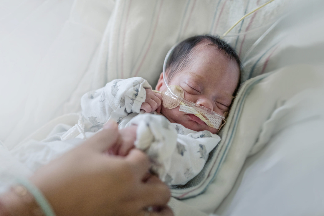
Brachial Plexus It’s a tough situation for any physician. After a difficult birth of a large baby, a limp, flaccid arm is noted and a brachial plexus injury is suspected. This injury occurs when the brachial plexus — a network of nerves that originate from the cervical spinal cord and supply axons to the skin and muscles of the shoulder, arm, and hand — is stretched.

Birth-related brachial plexus injuries are common, seen in 0.13 to 3.6 per 1,000 live births. Often, there is nothing that could have been done differently by physicians to prevent the injury. When families are told of the possible brachial plexus impairment, they are in disbelief, with many questions. How did this happen? What does it mean? Is my baby going to be normal? What are you going to do to fix it? Fortunately, infants with this type of injury can have good outcomes, but only if it is recognized early by the treating physician and the appropriate treatment is started.

“If kids don’t receive the early and proper treatment, you can’t go back in time and fix them,” says Gregory Heuer, MD, PhD, attending neurosurgeon at The Children’s Hospital of Philadelphia (CHOP). “The right treatment at the right time results in a good outcome.”

This outcome is greatly dependent on the degree of injury, the timing of surgery and, perhaps most importantly, whether or not the infant receives effective postnatal occupational therapy. CHOP is one of few institutions in the country with a comprehensive program to treat these children, with specialized services in Neonatology, Occupational Therapy, Neurology, Plastic Surgery, Neurosurgery, and Orthopedics.

After stabilization of the infant, our team first determines the extent of the injury. Brachial plexus injuries can range from mild, in which the axons remain intact (neuropraxia), to moderate, in which the axons are directly damaged (axonotomesis), to severe, in which the nerves are pulled out of the spinal cord or avulsed (neurotomesis). Factors associated with a poor prognosis also indicate a more severe injury. These include the presence of Horner’s syndrome, paralysis of the diaphragm, and total or lower plexus injury. Spontaneous recovery without surgery is seen in 70% to 95% of patients. This recovery occurs by 3 to 4 months of age and is complete by 12 to 18 months of age.

Our team obtains a complete obstetric history to look for risk factors associated with a plexus injury and attempts to document a complete motor and sensory exam. The infant is then examined to look for damage to nerves associated with the brachial plexus, specifically for paralysis of the diaphragm or for the presence of Horner’s syndrome. We look for factors that signal a difficult birth where a great deal of force was needed during the birthing process, including fractures of the clavicle or humerus, facial nerve injury, cephalohematoma, and torticollis. And we are on the lookout for signs or symptoms of other conditions that can mimic a brachial plexus injury, including orthopedic injuries, spinal cord injury, congenital varicella, lesions of the cervical spinal cord or brain stem, strokes, or tumors of the brachial plexus.

The next step in treatment is early initiation of occupational therapy. This normally begins 10 to 14 days after birth, allowing any pain,present after birth to subside. Therapy is focused on maintaining flexibility in the affected joints, particularly the shoulder, and monitoring and documenting changes in the motor exam of the infant.

If an infant fails to show signs of ongoing recovery, treatments are focused on returning function. These procedures can target reconstructing the nerves or the tendons, muscles, bones, or joints. The goal of nerve surgery is to improve motion and strength by supplying axons to power a muscle that lacks sufficient neural stimulation. In order to power a muscle and restore function, 30% of the original fibers are needed. This can be accomplished by resecting the scar tissue and placing nerve grafts to provide a path for new nerves to grow along. Nerve transfers are another technique of supplying axons by transferring a working nerve to stimulate the weak muscle. A combination of these methods, when done early enough, can result in significant improvement in the infant’s ability
to move. Depending on the muscle area that is weak, function improvement is seen in 60% to 90% of the individual muscle groups.

“Unlike most devastating conditions in a newborn, if these children are treated early enough, they can have a very good outcome,” says Heuer. “The potential to help them is tremendous.”

Some patients do not need nerve surgery or have residual weakness and/or stiffness after nerve surgery, but may be candidates for additional surgical procedures including muscle or tendon transfers, joint releases, osteotomies, and fusions. Muscle or tendon transfers take strong, working muscles to power weak muscle/tendon units. Joint releases are used for contractures and/or subluxations so that more motion can be obtained, and often may be done with muscle/tendon transfers. Osteotomies can reposition the arm or forearm such that it is in a more functional position, and fusions limit motion of the joint in order to provide more stability to the patient. These surgeries are sometimes done later in life, depending on the recovery of the child’s function. They often require a period of immobilization to allow for surgical healing and then postoperative therapy.

A. Anatomic relationship of the right brachial plexus. B. Schematic representation of nerves and branching of the brachial plexus. C. Shoulder dystocia: With child birth, traction can occur that stretches the brachial plexus. D. Nerve transfer: A nerve fascicle is transferred from a functioning nerve (bottom) to a damaged distal nerve target (top), supplying the muscle with a new, functioning proximal nerve to power it. E. Nerve repair: A damaged, scarred portion of a nerve, or neuroma, can be seen in the top image. This portion is resected, and, in the bottom image, nerve grafts are placed across the gap to repair the nerve.

Brachial plexus fast facts

  • Birth related brachial plexus injuries are common and need to be considered in the appropriate clinical setting.
  • Patients should be seen in the first few weeks after delivery at a comprehensive center for diagnosis and to start occupational therapy.
  • The natural history of patients with this condition is good, with 70% to 95% having a good outcome without surgery.
  • Surgery is directed at restoring function, either by supplying axons to the muscle or by altering the mechanics of affected muscles and joints. Although they may need multiple procedures, these children can have good functional outcomes.
Severity of Brachial Plexus Injury
InjuryTrade SeverityConditionRecovery
NeuropraxiaMildNerves stretched but remain in continuity and the axons remain intactSpontaneous recovery
AxonotomesisModerateNerves stretched and axons damaged and degenerate. Axon regrows from the spinal cord towards the target muscle at a rate of 1mm a day, unless blocked by scarVariable recovery – some require surgery
NeurotomesisSevereNerves can be pulled out of the spinal cord or avulsedNo recovery – surgery needed