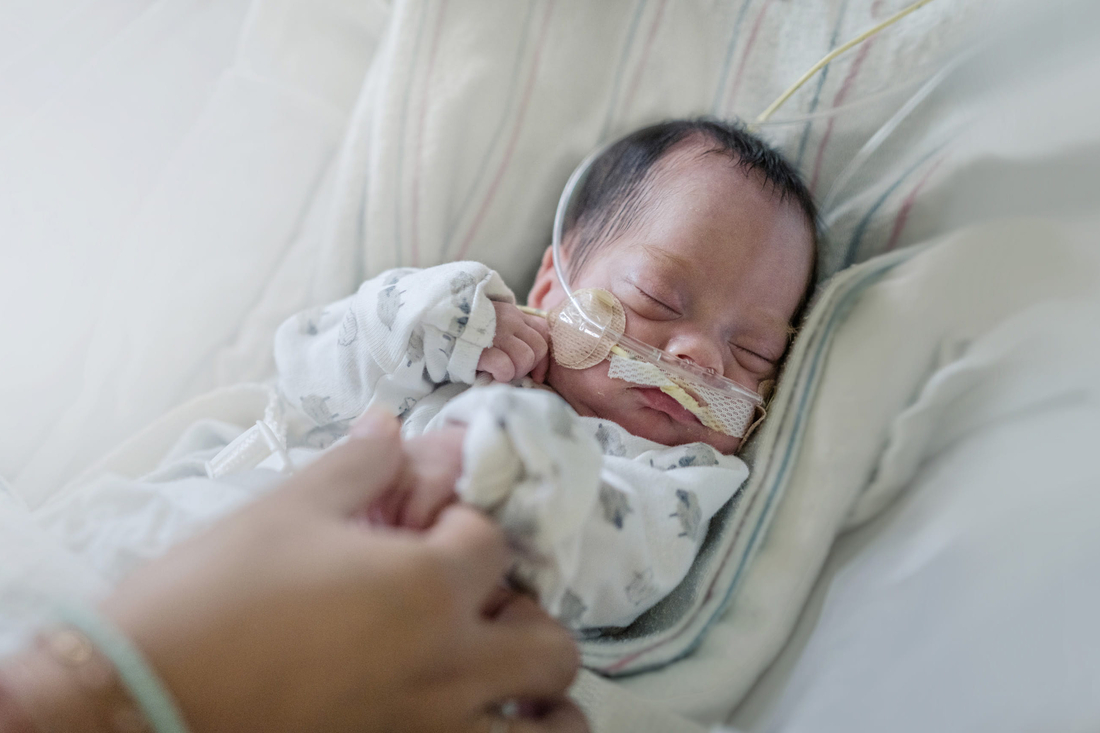
Division of Neonatology

Having a baby that needs intensive care is incredibly difficult. Your life may seem like it's been turned upside down. We are here for you at Children's Hospital of Philadelphia (CHOP). Our Neonatology team cares for very sick babies and those born early. We provide expert monitoring and care around the clock for all our patients. We help babies with many different serious illnesses. As part of one of the top children's hospitals in the United States, we can easily connect our patients to any other kind of medical help they might need, and we can perform all sorts of specialized surgeries for babies. In addition to our Philadelphia and King of Prussia inpatient locations, our neonatologists work at hospitals in the CHOP Care Network.
How we serve you
Your baby's neonatologist is not only an expert in providing specialized care, but is also involved in the development of new ways to treat these complex diseases more safely, effectively and with fewer side effects. Some of the most significant advances in neonatal care have happened right here at CHOP.
-
Breastfeeding and Lactation Program -
Infant Breathing Disorder Center -
Neonatal Airway Program -
Neonatal Craniofacial Program -
Neonatal Follow-up Program -
Neonatal Neurocritical Care Program -
Neonatal Surgical Team -
Newborn & Infant Chronic Lung Disease Program -
Newborn/Infant Intensive Care Unit at CHOP -
Resilience After Infant Substance Exposure Program

Why choose CHOP for neonatal care
We are world-recognized leaders in caring for critically ill infants. Here, you and your baby benefit from the expertise of our vast team, our strong history of innovation and our family-focused care.

Meet your team
Here, your baby's care is in the hands of one of the largest, most accomplished teams of neonatal experts in the world, including physicians, advanced practice nurses, physician assistants, registered nurses, developmental therapists and more.

Our locations
Access CHOP neonatal care in one of our many locations throughout the region.

Our research
The Division of Neonatology has a robust research program. We are leading research to increase our understanding of diseases in babies and improve treatment of sick babies.

Resources for families
We know this is an emotional and stressful time for your family. We have pulled together resources to help you find answers to your questions and feel confident with the care you're providing your baby.

Resources for professionals
Everything you need to support your patient’s health, created and updated by our CHOP community of experts.
Events
4th Annual Lymphatic Conference
Thursday, Sep 18, 2025
Your donation changes lives
A gift of any size helps us make lifesaving breakthroughs for children everywhere.


