What is bronchopulmonary sequestration?
Bronchopulmonary sequestration, also known as BPS or pulmonary sequestration, is a rare birth defect in which an abnormal mass of nonfunctioning lung tissue forms during prenatal development. It can form outside (extralobar) or inside (intralobar) the lungs, but is not connected directly to the airways.
The abnormal lung tissue does not function like normal lung tissue. These masses are characterized by abnormal blood supply, in which a systemic arterial blood vessel coming from the aorta feeds the lung mass.
Treatment for bronchopulmonary sequestration depends on the type and size of lung lesion, as well as whether the condition is causing any serious health complications for mother or baby.
While some cases of small extralobar BPSs will not require surgery, large extralobar BPSs and all intralobar BPSs can lead to breathing problems, infection, and life-threatening complications like heart failure. Surgery is needed to remove the abnormal tissue.
Most children with bronchopulmonary sequestration can be safely treated with surgery after birth. In rare cases — when the lesion has grown abnormally large, is restricting lung growth or impairing blood flow, putting your baby at risk for heart failure — fetal intervention may be necessary.
Types of BPS
There are two types of bronchopulmonary sequestration:
- Intralobar, in which the mass forms inside the lungs. These lesions account for about 75% of cases of BPS, affect males and females equally, and are generally isolated birth defects. All intralobar lesions require surgical removal (resection) after birth.
- Extralobar, in which the abnormal mass forms outside — but nearby — the lungs. In some instances, this type of BPS may be located in the abdomen. These lesions account for only about 25% of BPS cases, and are more likely to affect males than females. Small extralobar BPS can frequently be managed without surgery after birth, while large lesions will require surgery.
Causes of BPS
The cause of bronchopulmonary sequestration remains unknown. It has not been linked to a genetic or chromosomal anomaly, and does not appear to run in families (is not hereditary).
Most clinicians believe the condition begins during prenatal development when an extra lung bud forms and migrates with the esophagus. Depending on when the extra lung bud forms, it may become part of one of the lungs (intralobar), or grow separately (extralobar).
Signs and symptoms of BPS
Symptoms of bronchopulmonary sequestration can vary, and depend on the size of the lesion.
After birth, children with BPS may experience:
- No symptoms
- Trouble breathing
- Wheezing or shortness of breath
- Frequent lung infections like pneumonia
- Upper respiratory infections that take longer than usual to resolve
- Feeding difficulties and trouble gaining weight as infants
All suspected lung lesions, whether found before or after birth, require careful imaging. Determining the type, size, and location of the lesion will guide treatment recommendations.
Evaluation and diagnosis of BPS
Bronchopulmonary sequestration is one of several types of congenital lung lesions and may be confused with congenital cystic adenomatoid malformation (CCAM). While similar in some ways, BPS and CCAM are unique conditions that require individualized treatment. A child can also develop a hybrid lesion, which has characteristics of both a BPS and CCAM. This unusual condition makes diagnosis challenging.
Thanks to improvements in prenatal imaging, most cases of BPS are discovered during routine ultrasounds between 18 to 20 weeks' gestation. A solid mass will typically appear on the ultrasound as a bright spot in the fetus’s chest cavity. Expert fetal imaging specialists experienced in evaluating fetal lung lesions can detect the source of the blood flow to the lung lesion as well how blood is drained from the lesion. This is an important step to confirm an accurate diagnosis and distinguish between an intralobar and extralobar BPS, hybrid lesion, CCAM, or other type of fetal lung lesions.
At Children’s Hospital of Philadelphia (CHOP), our fetal radiologists, maternal-fetal medicine specialists and fetal surgeons have established ways to distinguish between the different types of lesions before your baby is born. This accurate diagnosis helps us create an optimal plan for the care of your child after birth. (Read a paper published by our team that established these diagnostic standards.)
If you are carrying a baby suspected to have BPS, you should be seen by a center with expertise in lung lesions for a more thorough examination. More than 3,013 patients with suspected lung lesions have been referred to our team — this experience makes us uniquely equipped to evaluate and manage your baby’s condition.
When you come to the Center for Fetal Diagnosis and Treatment, you will have a comprehensive one-day evaluation. Our fetal imaging specialists use the most advanced prenatal imaging techniques available to gather detailed information about your baby’s health and make an accurate diagnosis. In most cases, you will have a high-resolution fetal ultrasound, fetal echocardiogram to evaluate heart structure and function, and in some cases an ultrafast fetal MRI (developed at CHOP).
These specialized tests help us determine the type, size and location of the mass, where the blood supply is coming from, and allow us to see any impact the mass may be having on your baby’s heart.

Why choose CHOP for lung lesion care
CHOP provides comprehensive care for both mother and baby diagnosed with fetal lung lesions. From before birth to long-term follow-up, we are here for you every step of the way.
Management of pregnancy with BPS
Depending on the gestational age of your baby and the size of the mass, you will continue to have regular ultrasounds to closely monitor the growth of the lung lesion.
Rarely, these masses can grow quite large, taking up valuable space in the chest. This can restrict normal lung growth and can lead to underdeveloped lungs which will not function adequately at birth. Large masses can also shift the heart and impair blood flow. This can lead to fetal heart failure (fetal hydrops) and cause the buildup of fluid in the fetus and placenta.
Some of these masses are associated with a large pleural effusion, or fluid collection in the chest cavity. This fluid collection can also compromise the ability of the fetal heart to function normally.
Over several visits, clinicians will determine how quickly your child’s BPS is growing.
Fetal intervention for BPS
Treatment for bronchopulmonary sequestration depends on the type and size of lung lesion, as well as whether the condition is causing any serious health complications.
Some babies with bronchopulmonary sequestration cannot wait for treatment after birth because the lesion is too large, growing too rapidly, or causing life-threatening complications in utero such as fetal heart failure.
Fetal interventions we offer to treat bronchopulmonary sequestration include:
Draining fluid from the chest
A small number of bronchopulmonary sequestrations can develop a large pleural effusion, or accumulation of fluid in the chest, outside of the lung, which can compress the lungs and heart. This fluid can be drained prenatally and a shunt can be left in place to provide continued drainage of the fluid.
The shunting procedure itself is performed under ultrasound guidance. A large trocar (hollow needle) is guided through the mother’s abdomen and uterus, and into the fetal chest. The shunt is passed through the trocar to divert the accumulated fluid from the fetal chest to the amniotic sac. The shunt will remain until delivery. The goal of these procedures is to decrease the accumulation of fluid to ward off heart failure (fetal hydrops).
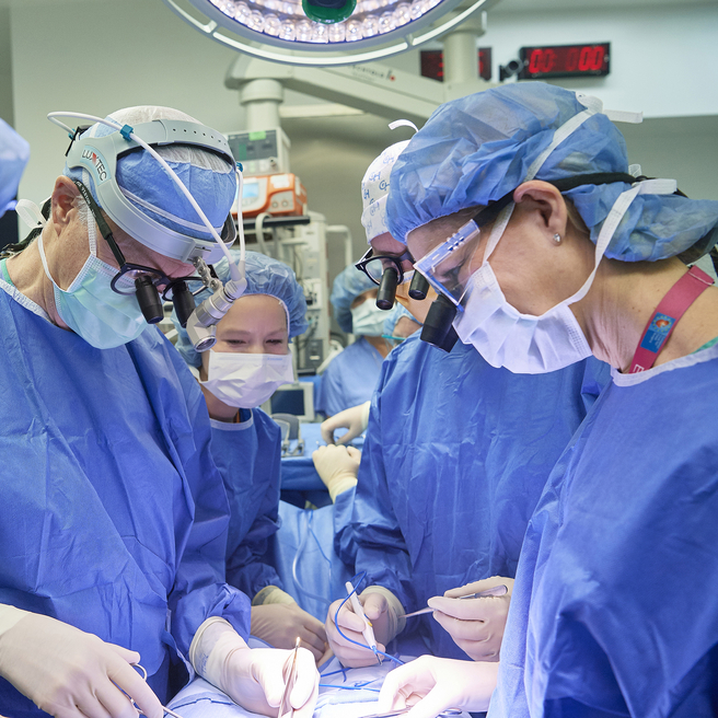
C-section to resection
Babies with large lung lesions can be safely delivered by c-section and be carried immediately to the adjacent operating room where our expert fetal surgeons will remove the mass. Our Garbose Family Special Delivery Unit (SDU) was specifically designed with adjacent fetal operating rooms for this purpose. After the mass is removed, our dedicated Neonatal Surgical Team will provide further specialized care for your baby.
EXIT procedure
Rarely, a large BPS may require a specialized delivery technique, such as the ex utero intrapartum treatment (EXIT) procedure. The EXIT procedure is performed in our SDU.
In an EXIT procedure, your surgical team will partially deliver the baby so that they are still attached to the placenta and receiving oxygen through the umbilical cord. This procedure allows time for fetal surgeons to establish an airway and remove the mass while the baby is still attached and supported by the mother. After removal, your baby will be delivered and our Neonatal Surgical Team will provide further specialized care.
Delivery of babies with BPS
Mothers carrying babies with small lung lesions — without other associated anomalies — may be able to deliver at their local hospital, without the need for high-risk neonatal care. Babies with larger lesions, or those with complications or associated disorders, should be delivered in a center that offers expert care for both mother and baby in one location.
At CHOP, babies with prenatally diagnosed lung lesions who will require treatment immediately or soon after birth are delivered in the Garbose Family Special Delivery Unit, specifically designed to keep mother and baby together and avoid transport of fragile infants.
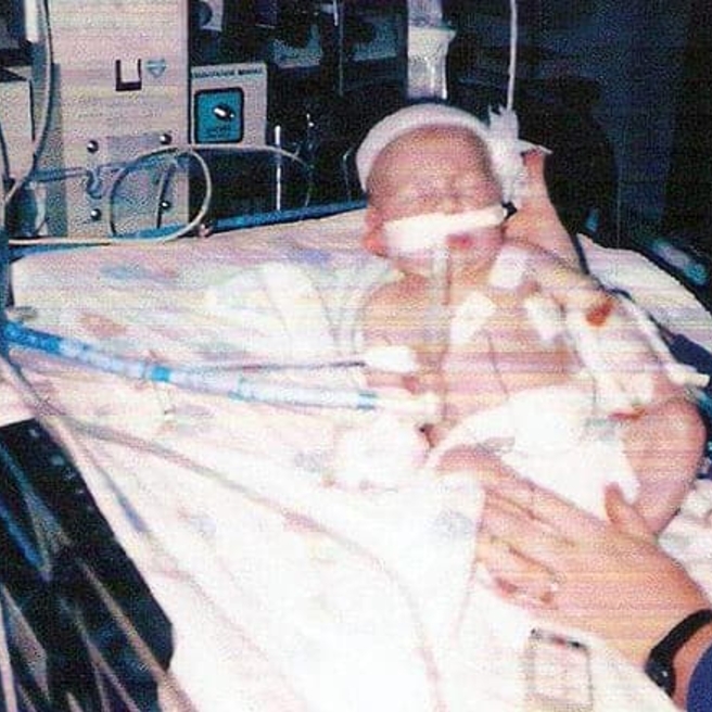
Here, your baby has immediate access to the Newborn/Infant Intensive Care Unit (N/IICU) and a dedicated Neonatal Surgical Team. Our team is experienced in performing complex, delicate procedures needed to establish an airway while delivering babies who may not be able to breathe on their own at birth, as well as any immediate surgeries that your baby might need.
Surgery for bronchopulmonary sequestration after birth
BPS lesions can be successfully treated with surgery after birth.
- All intralobar BPS lesions should be surgically removed because of an increased risk of infection as well as the potential for high blood flow through the tissue that can lead to heart failure later in life.
- Large extralobar BPS, especially those with high blood flow, may compromise your baby’s ability to breathe or put too much stress on your baby’s heart, and should be surgically removed.
- Small extralobar BPS may not require surgery to remove the lesion.
Removing the BPS mass when your child is young has multiple benefits, including promoting compensatory lung growth (ability of lungs to grow and fill the space in the chest) and avoiding potential complications such as lung infections.
First, you will come in for an appointment for your child to be evaluated by the surgical team. A CT scan with contrast will be performed to confirm the diagnosis and determine the exact location of your child’s lung lesion. Children’s Hospital of Philadelphia radiologists have set the standard for safe, low-dose imaging practices in children. We make every effort to schedule surgery as soon as possible and at a time convenient for your family. Surgery is often scheduled for the same week.
The average length of stay at CHOP after lung lesion surgery is two to three days.
Follow-up care
Follow-up care for children with bronchopulmonary sequestration will depend on the treatment the child received.
Most children treated for small lesions after birth will only need monitoring for the first year after surgery to ensure normal lung growth and lung function. The majority will require no additional long-term follow-up care.
Children treated for more severe or complex lung lesions may require ongoing monitoring through childhood. For those who experience limited lung growth resulting in pulmonary hypoplasia, CHOP’s unique Pulmonary Hypoplasia Program (PHP) offers comprehensive long-term care. The PHP team provides multidisciplinary care with a focus on improving your child’s pulmonary health, evaluating neurodevelopmental growth, meeting nutritional needs, monitoring for late onset hearing loss or any surgical issues, and more.
Long-term outlook
The vast majority of children with congenital lung lesions, including BPS, do extremely well and have normal lung function after their lesions are removed. This is due to rapid compensatory lung growth that occurs during childhood. Having surgery early maximizes this compensatory growth.
Children with moderate to large lesions can also do extremely well, but their outlook depends on expert treatment to avoid potential complications. These babies require highly specialized expert care from time of diagnosis to delivery and surgery to ensure the best possible long-term outcomes.

Tour our Fetal Center
The Wood Center for Fetal Diagnosis and Treatment has cared for many families and will help you through your journey, too.

What to expect
From the moment of referral through delivery and postnatal care, your family can expect a supportive experience when you come to us with a diagnosis of a birth defect.
Resources to help
Bronchopulmonary Sequestration (BPS) Resources
Richard D. Wood Jr. Center for Fetal Diagnosis and Treatment Resources
Learning your baby has a birth defect is a life-changing experience. We want you to know that you are not alone. To help you find answers to your questions, we've created this list of educational health resources.
Meet your team
You will receive care from a highly skilled and compassionate team with a global reputation. This team helped shape modern fetal care and continues to improve techniques and treatments.
Patient stories
Reviewed by N. Scott Adzick, MD, MMM, FACS, FAAP