Cancer Center

When your child is diagnosed with cancer, it can be overwhelming. You and your child might experience many emotions, such as feeling scared and confused.
Our pediatric cancer team at Children's Hospital of Philadelphia (CHOP) is here to guide you through your child's treatment journey. Our internationally known oncologists have expertise in every type of cancer, from the rarest to the most common. We offer the widest possible range of treatment options, including clinical trials only available here. Your child will receive a specialized, comprehensive care plan.
We also provide a level of support to families that is not always available in other centers. Our more than 175 team members will meet any need your family may have.
How we serve you
To give your child the very best care, our doctors, nurses and other experts are grouped into specially trained treatment teams. These teams provide your child with personalized care while building strong relationships to support your family.
-
Advanced Pediatric Thyroid Cancer Therapeutics Clinic -
Alex Scott Day Hospital -
Apheresis Program -
AYA Oncology Program -
Blood and Marrow Transplant Program -
Bone and Soft Tissue Tumor Program -
Brain Tumor Program -
Cancer Predisposition Program -
Cancer Survivorship Program -
Cellular Therapy and Transplant -
Comprehensive Vascular Anomalies Program (CVAP) -
Developmental Therapeutics Program -
ELECT Program -
Executive Function Clinic -
Fertility Preservation Program -
Financial Assistance -
Home Care Program -
Immunotherapy Program -
Integrative Oncology Program -
Leukemia and Lymphoma Program -
Oncology Neuropsychology Screening Program -
Oncology Psychosocial Services Program -
PHTS Clinic -
Physical Therapy Department -
Precision Medicine for High-Risk Pediatric Cancer -
Proton Therapy Center -
Refractory Neuroblastoma Program -
Relapsed Leukemia and Lymphoma Program -
Revaccination Clinic -
Solid Tumor Program -
Surgical Oncology Program -
Urologic Oncology -
Very Rare Malignant Tumors Program
Conditions we treat
We treat every type of childhood cancer, including cancers that are very rare.
-
Acute lymphoblastic leukemia (ALL) -
Acute myelogenous leukemia (AML) -
Brain tumors -
Central nervous system (CNS) germ cell tumors -
Chondrosarcoma -
Chronic myelogenous leukemia (CML) -
Differentiated thyroid cancer -
Ewing sarcoma -
Hepatoblastoma (liver cancer) -
Hodgkin lymphoma -
Juvenile myelomonocytic leukemia (JMML) -
Leukemias -
Managing mucositis -
Medullary thyroid cancer -
Mucopolysaccharidosis type 1 (MPS 1) -
Mucopolysaccharidosis type II (MPS II) -
Neuroblastoma -
Non-Hodgkin lymphoma -
Osteosarcoma (bone cancer) -
Relapsed or refractory neuroblastoma -
Relapsed or refractory acute lymphoblastic leukemia (ALL) -
Retinoblastoma (eye cancer) -
Rhabdomyosarcoma -
Skin cancer -
Soft tissue sarcomas -
Testicular tumors -
Thyroid cancer -
Wilms Tumor (kidney tumor) -
Wiskott-Aldrich syndrome (WAS)

Why choose us for cancer treatment
Our large program offers cutting-edge treatments and services that aren't available anywhere else. We are ranked among the top pediatric cancer centers in the nation by U.S. News & World Report and Newsweek. Some of the biggest breakthroughs in the treatment of pediatric cancers have happened right here.

Meet your team
Your child will be cared for by one of the most accomplished teams of childhood cancer experts in the world. We provide medical care, emotional counseling and much more. We make sure your child, you and every family member get the highest level of support.

Cancer Center locations
Access top pediatric oncologists who treat children with cancer.

Our research
Our team of multidisciplinary specialists at the Center for Childhood Cancer Research is dedicated to decreasing side effects and improving the cure rates of pediatric cancers with a diverse collection of translational and clinical research initiatives.

Cancer Center resources
We have created resources to help you find answers to your questions and feel confident with the care you are providing your child.

Resources for professionals
Everything you need to support your patient’s health, created and updated by our CHOP community of experts.
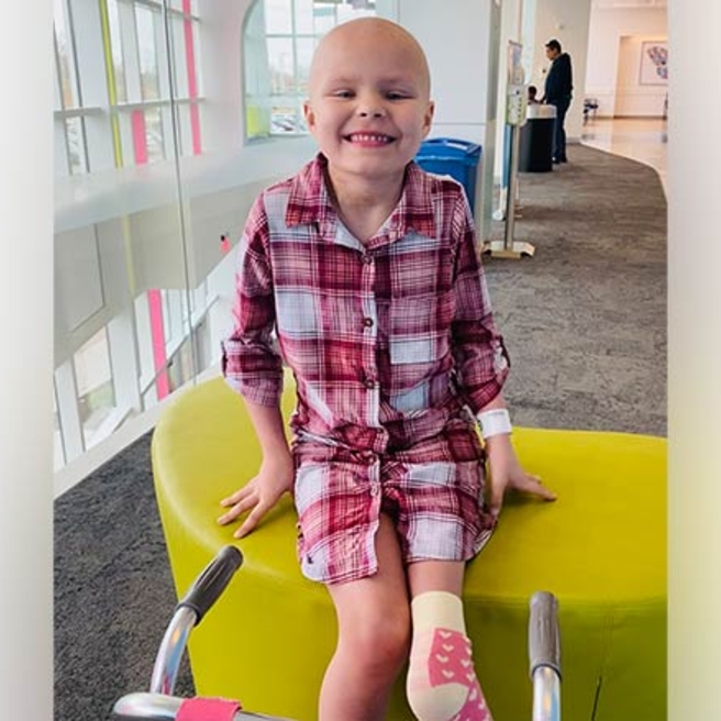
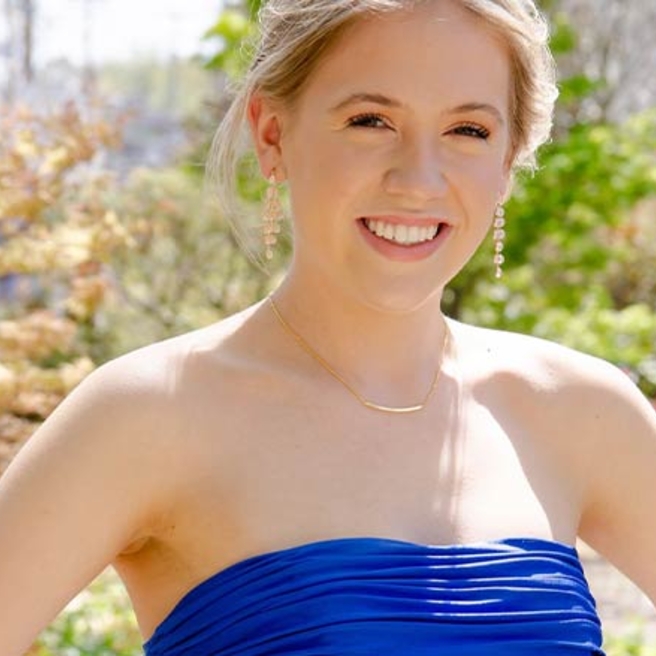
Patient stories
Events

Parkway Run & Walk, presented by Citadel Credit Union
Sunday, Sep 28, 2025

Connections: CHOP/Momcology Group for Caregivers - Nov 15
Saturday, Nov 15, 2025

Audrey's Children
A new biopic shares the untold true story of renowned CHOP oncologist, Dr. Audrey Evans, an oncology pioneer and trailblazer for women in medicine.
Give to the Cancer Center
There are many ways to support CHOP’s Cancer Center. A gift of any size can help advance our work for children everywhere.
