Fetal Surgery Experience at Children's Hospital of Philadelphia - Online Course

Transcript

N. Scott Adzick: Hi. I'm Scott Adzick. I serve as the surgeon in chief at The Children's Hospital of Philadelphia and the director for the Center for Fetal Diagnosis and Treatment at CHOP. We're going to have some fun today and review some elements of the fetal diagnosis and therapy experience at The Children's Hospital of Philadelphia.
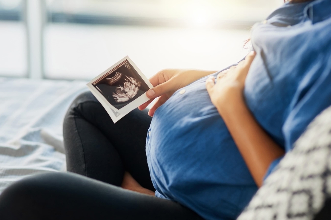
Here's the real star of the show. When you think about it, until about 25 or 30 years ago, the human fetus was a very mysterious little creature hidden away inside the mom as a medical recluse. But over the past three decades, due to advances in prenatal diagnosis, we've learned that the fetus can have serious medical problems and may even need our help.

And I'd like to try to convince you that Samuel Taylor Coleridge was right when he wrote 200 years ago, "The history of man for the nine months preceding his birth would probably be far more interesting and contain events of greater moment than all of the threescore and ten years that follow it."

I have a short video to show which puts the context of what things were like 30 years ago when we first started to consider the repair of life-threatening birth defects before birth.

For most of human history the fetus has been really shrouded in mystery and multiple layers of the maternal abdomen obscuring anything that was wrong with the fetus.

With the emergence of prenatal diagnostic techniques, particularly with the advent of maternal-fetal ultrasound, we could go into the womb for the first time.

We could see things we’d never seen before, and as those fetuses were followed, it became more clear what may or may not happen to them.

There were a certain category of birth defects that were, for the most part, lethal, where when the baby was born we were too late to do anything about it.

That was really the impetus to try to knock on the door sooner.

Perhaps we can treat this new group of highly selected, unborn patients who have severe diseases that are progressive during the pregnancy. Maybe we could treat them before birth, and that was a very radical and controversial concept.

Here is a summation of the problem of birth defects. They're common—one in 28 babies is born with a structural birth defect; costly—billions of dollars are required for medical treatment; merciless—no parent is immune; mysterious—most causes of birth defects are unknown; overlooked, in my opinion—the research is under-funded; and deadly—birth defects are the leading cause of infant mortality.

Prenatal therapy requires, of course, prenatal diagnosis, whether it be pregnancy screening, high-resolution ultrasound, fetal MRI—which we played a role in developing for looking at birth defects before birth—fetoscopy, molecular diagnosis, the effects of the Human Genome Project, to the point where now most human anatomic and genetic abnormalities are diagnosed before birth, and the question is can we treat before birth?

Now, as a pediatric surgeon, in my day-to-day life I counsel many mothers carrying babies with birth defects. And the prospect of an abnormal fetus permits a variety of management alternatives, including abortion. The birth defect may mandate a change in the timing of delivery, preterm versus term, a change in the mode of delivery, vaginal delivery versus Cesarean section. It may change how the delivery is performed, as I'll describe. Ex utero intrapartum therapy is used in highly selected circumstances at the delivery time. In most cases we can wait until near term or term, transport the baby inside the mom, maternal transport, as opposed to the baby being born elsewhere and then being transported to CHOP by helicopter or by airplane or by ambulance, and then do diagnosis and therapy after birth. And then there are a few uncommon birth defects where it makes sense to fix them before birth.

We decided many years ago, when Mike Harrison and I were working together at University of California San Francisco, that open fetal surgery is only justifiable if there is a poor prognosis without intervention, if it's been shown to be efficacious in animal models, and if all this can be done at relatively low maternal risk, keeping in mind that the mother is an innocent bystander medically.

So we can now diagnose fetal diseases by ultrasound and other techniques. We then go to the laboratory and in animal models, principally fetal sheep and fetal monkeys, create models of birth defects, study the pathophysiology, and determine whether an in utero correction can end up with a good kid. And all this needs to be done—all this homework needs to be done prior to applying it clinically.

We did experiments in literally thousands of fetal sheep and in 400 or so fetal rhesus monkeys at the California Primate Research Center in Davis, California, where we developed the surgical anesthetic and tocolytic techniques to safely do fetal surgery in the non-human primate model. We showed that, compared to the controlled breeding colony, there was no impairment of future reproductive capacity and worked on techniques to minimize the risk of preterm labor which, of course, is still an unsolved problem.

This shows the schematic view of what a mother goes through and the fetus goes through in an open fetal surgery operation, and I'll describe this in greater detail as well as show some video clips of open fetal surgery procedures.

Here's a list of anatomic defects on the left-hand side and the effect on development. And I'm going to touch on most of these, keeping in mind that there are CME credit lectures on the CHOP website for some of these lesions where we go into great detail about indications for therapy and so forth. Fetal lung lesions is one of those CME lectures which I'll touch on. Large lesions can result in pulmonary hypoplasia and fetal hydrops. Sacrococcygeal teratoma that can cause high output cardiac failure with very large solid lesions. Causes of fetal airway obstruction, laryngeal atresia, or lesions in the neck, like a cervical teratoma, can mandate a change in how the baby is delivered, using the ex utero intrapartum therapy strategy, or EXIT procedure. I'll touch on twin-twin transfusion and TRAP syndromes. Dr. Mark Johnson has a CME lecture in which this is discussed in detail. Myelomeningocele I'll touch on—but there's also a very detailed lecture on this that I have given for CME credits on fetal surgery for spina bifida trials and tribulations. Urinary tract obstruction, congenital diaphragmatic hernia, and certain causes of—certain types of heart disease diagnosed before birth.

To do this sort of work requires a team, and we have an incredible team in the Center for Fetal Diagnosis and Treatment at CHOP. Lori Howell is the executive director. We have talented pediatric surgery faculty at CHOP. We have 75 full-time pediatric surgical specialists. In any sort of specialty one can think of, we can bring all that expertise to bear on the baby with a prenatally diagnosed birth defect. We have four full-time obstetricians, maternal-fetal medicine specialists, Drs. Johnson, Moldenhauer, Khalek, and Martinez-Poyer, as well as five OB generalists who run our Special Delivery Unit, talented neonatologists—and I mention Natalie Rintoul here—experts in fetal imaging, Beverly Coleman for maternal-fetal ultrasound, Jack Rychik for fetal echocardiography, and Ann Johnson, Tedi Victoria, and the neuroradiologists for fetal imaging using ultra-fast fetal MRI techniques.

Anesthesiologists, geneticists, nurses. A real strength is our mental health services program for mothers who go through a lot of stress in terms of dealing with the consequences of bearing a baby with a birth defect. And the Special Delivery Unit, operating room and N/IICU staff, of course.

This shows our considerable clinical experience. Since the program started in 1995, more than 15,000 referrals, more than 1,000 fetal surgery procedures, close to 100 EXIT procedures, referrals from all 50 states and from more than 50 countries outside the U.S.

Referral diagnoses during that time period are shown here, more than a thousand fetal lung lesions, nearly a thousand congenital diaphragmatic hernias, many twin-twin transfusion and the like diagnoses, and so forth.

We have the asset at The Children's Hospital of Philadelphia of the Garbose Family Special Delivery Unit, which is a state-of-the-art unit totally focused on the mother carrying a baby with a birth defect. The Garbose family generously supported this unit, which is quite unique. That's Lynne Garbose there in the middle with a baby in her arms, and next to her is a mother who underwent fetal surgery for spina bifida weeks before. In this unit, there's the inpatient care for labor and delivery, we have custom-made fetal surgery operating rooms and neonatal resuscitation rooms, and the outpatient portion of the Special Delivery Unit has facilities for ultrasound, for echocardiography, and for MRI.

I gave a very detailed CME lecture on the CHOP website on fetal lung lesions, along with my colleague, Dr. Alan Flake, so I'll just touch on some of the elements for fetal lung lesions in this lecture. There's a broad spectrum of clinical severity which we've learned from following 1,300 or so babies with prenatally diagnosed lung lesions. Small to moderate-sized lesions can be followed during the pregnancy and dealt with after birth. Very large lesions, as shown in this schematic view with a lesion arising from the left lower lobe, can cause a big mediastinal shift, esophageal compression, polyhydramnios, and because it's a large space-occupying lesion, hypoplasia of the normal lung tissue, as well as compression of the vena cava and heart, leading to fetal cardiovascular failure or fetal hydrops, characterized by the accumulation of fluid within the body cavities of the fetus.

This is straightforward to diagnose before birth, and the most common type is a congenital cystic adenomatoid malformation, or a CCAM or CCAM for short. This shows an ultrasound view in the middle of a fairly substantially-sized CCAM lesion, and on the right side is a cystic and solid CCAM shown by fetal MRI. Years ago, we characterized CCAMs into two categories, macrocystic and microcystic. One can get additional anatomic information by fetal MRI, and the prognosis for the fetus relates principally to the growth and overall size of the lesion, which is more important than the stock or histologic type of CCAM.

The pathognomonic finding of a bronchopulmonary sequestration is shown here by color flow Doppler ultrasound showing a large systemic arterial feeding vessel coming off the thoracic aorta of the fetus. That's the hallmark pathognomonic finding for a bronchopulmonary sequestration. And of course, there can be lesions that are both a CCAM and a sequestration, and we coined the term hybrid lesion years ago.

The CCAM mass volume/head circumference ratio, or CVR, is a measurement pioneered at CHOP by Dr. Beverly Coleman and Dr. Tim Crombleholme more than a decade ago. We followed the growth profiles of a large number of lesions and have learned that those with a CVR greater than 1.6 are at much higher risk than those with smaller lesions. We've also shown that in lesions not developing fetal hydrops, rapid growth is possible between 18 to 25 weeks gestation. But usually there's a growth plateau at 25 to 28 weeks gestation, and oft times there is no growth after that plateau period except for some large cystic lesions. And regression in size of the lesion latent gestation is fairly common, and in some cases, the lesion becomes isoechoic with the normal lung tissue and can be difficult or impossible to see.

The CCAM volume ratio, or CVR, is useful in terms of sonographic surveillance. For those patients with a CVR greater than 1.6, they're at risk for hydrops and warrant twice weekly evaluation. For those in the mid-range of 1.2 to 1.6 can be seen initially twice weekly and then weekly thereafter, depending on growth. And those with a CCAM volume ratio of less than 1.2 can be followed less frequently.

We and others have shown that maternal betamethasone therapy for congenital cystic adenomatoid malformation is useful for those lesions that are principally solid, and had a publication about this about six years ago. Betamethasone is ineffective for macrocystic lesions. We currently recommend this benign therapy for fetal CCAM cases with a CVR greater than 1.4. And this mode of therapy, which likely stunts the growth of the lesion, has led to a dramatic decrease in the need for in utero CCAM resections or thoracoamniotic shunt placements as a result.

For those fetuses with huge CCAM lesions or bronchopulmonary sequestrations where the fetus develops hydrops, the mother can become sick with what we've coined as the maternal "mirror" syndrome, which is basically a preeclamptic state characterized by placentomegaly, a sick, water-logged placenta due to the fetal cardiovascular failure. This preeclamptic state may be characterized by maternal hypertension, proteinuria, peripheral and pulmonary edema. These mothers may require a transfer to the intensive care unit. It's been seen in other causes of placentomegaly may be related to release of placental vasoactive factors or an endothelial cell toxin and can be reversed only by delivery of the sick fetus and placenta. And invariably, the sick, hydropic, premature baby dies.

So we decided years ago that it might make sense to go after to treat those CCAM lesions in which the fetus develops hydrops but the mother has not yet developed the maternal mirror syndrome. Working in Mike Harrison's lab 25, 30 years ago, I developed techniques for doing fetal lobectomies and pneumonectomies. And this shows a mid-gestation lamb who was undergone a thoracotomy and is about to undergo a lobectomy.

This shows our first successful fetal CCAM resection case and this was done at 22 weeks gestation. Here he is as an infant destined to be a baseball player. In the middle he is a T-ball player and on the far right he is throwing out the first pitch at a Phillies’ spring training game at age 16 and his ambition is to be a college baseball player.

And we show that after lobectomy or pneumonectomy there’s remarkable compensatory lung growth of the remaining lung tissue.

This shows a characteristic CCAM case at 21 weeks gestation. On the far left, a CVR of 2.0 because this was principally solid, the fetus was treated with steroids 13 days thereafter. This large left lower lobe lesion, which is shown by MRI in this case, failed to have a rest of the growth of the lesion with maternal steroids. There was integral growth over the next two weeks, which is huge in cross sectional view on the MRI and the middle lower part of the slide. By that time, the CVR was up to 3.3 with hydrops including ascites, scalp edema, and echocardiographic findings consistent with cardiovascular failure so we did an in utero resection at 23 weeks gestation.

This shows a video clip of that operation at 23 weeks gestation. The uterus is exposed. There’s an anterior placenta, so we worked posterior fundal. Full thickness stay sutures replaced, the Bovie cautery is used to incise down through the myometrium and the membranes. The uterine stapling device, that we helped to invent, is then applied. That’s very hemostatic. The left arm is exposed, as well as the left chest. Intravenous access is obtained for medications or blood transfusion. A miniaturized pulse oximeter is placed then wrapped in foil to prevent interference with light. The left thoracotomy is performed. You see us going between the ribs here to expose a huge left lower lobe lesion, which is coming into view. It is gently removed from the chest through a very large thoracotomy because there is a tamponade effect and we want to make sure by fetal echocardiographic monitoring that the fetus has adequate preload particularly once the lesion is decompressed. The fetal thoracotomy is closed and here is the uterus being closed in two layers.

This shows schematically on the left at 23 weeks gestation, huge left lower lobe lesion, the signs of fetal hydrops with ascites, skin and scalp edema. Six weeks post-op, at 29 weeks gestation, the heart is back in the midline, there is remarkable compensatory lung growth and usually within 10 to 14 days the signs of fetal hydrops resolves.

And this shows another child who underwent resection at 24 weeks gestation.

Dr. Mark Johnson and his colleagues are experts at placing thoracoamniotic shunts to decompress large cystic lesions associated with hydrops. And this shows the instruments for fetal shunt placement.

This is a double pigtail catheter that is placed under sonographic guidance with one end stretched out through a trocar and placed into the largest cystic portion of the lesion. And then the trocar is pulled back, the straight portion of the catheter goes to the chest wall. And then the other pigtail is in the amniotic fluid which allows fixation and for decompression of the large cystic lesion into the amniotic fluid.

This shows a macrocystic CCAM at 24 weeks gestation with fetal hydrops. You could see the huge lesion, massive ascites, massive skin and scalp edema, a CCAM volume ratio of 3.6, and a thoracoamniotic shunt is placed. And in a transverse view by ultrasound you can see the arrow showing the pleural amniotic shunt. The CVR is down to 0.9 after the shunt was placed.

And this is what the shunt looks like at the time of birth.

We have a large experience with ex utero intrapartum therapy, or the EXIT procedure, to resect chest masses that are very large latent gestation; 24 cases, with 21 survivors.

Many of these cases have not only very large CVR's late in gestation, but also at presentation. 17 of the 24 had fetal hydrops, 16 had polyhydramnios, many had both. 16 of the 24 required fetal thoracentesis, or shunt placement, or amnioreduction, and/or betamethasone.

The time on placental bypass was 65 minutes, with a range of 50 to 82 minutes. Four of the 21 survivors required the use of ECMO within 24 hours for pulmonary hypertension. Maternal complications included preterm labor in five cases due to polyhydramnios, chorioamnionitis in one, and a maternal blood transfusion in one case.

Postoperative complications included an air leak that required a reoperation, postoperative chylothorax which resolved on its own, and there were 3 deaths. One was a reoperation for bleeding and death in the 27 week gestation newborn, which was a desperate situation. Pulmonary hyperplasia and use of ECMO after a shunt and steroids in a baby who was treated before birth and managed with the ECMO approach and with ECMO after birth, but died of pulmonary hypoplasia death. And then a second patient who had pulmonary hypoplasia even after shunt placement and steroid courses times three.

This shows a case with a CVR of 2.6, and fetal hydrops, received steroids. The lesion stayed stabilized at 27 weeks gestation, but at 37 weeks gestation there was still a persistent ascites, a solid mass effect, and an EXIT approach was then used.

This video shows the EXIT approach. This is done through a low transverse uterine segment hysterotomy using the uterine stamping device. The fetal head, neck, and chest is delivered. The fetus is anesthetized through deep general anesthesia for the mother and with an additional injection of muscle relaxant and narcotic. The fetal chest is then exposed. A thoracotomy is performed. There is this very large lesion that's present. And the point is to decompress this and to resect this during the EXIT operation to allow full expansion of the normal lung tissue. The feeding and draining vessels and the bronchus is elongated and divided, and the lesion is then removed. So the fetus is still on maternal bypass through the umbilical cord and the placenta.

Here's the lesion being removed. That's the lesion. Here's the chest being closed. The bottom half of the fetus is still inside the mom. We oft times then will ventilate, if need be, instill surfactant, and then once we have control of the umbilical vessels and place a UA and possibly a UV line, and then clamp and divide the cord and deliver the baby who goes next door to a neonatal resuscitation room.

This shows our approach with lung lesions in terms of prenatal management. Whether it be a CCAM, a hybrid lesion, or an intralobar or extralobar BPS, the key feature is whether there is a mass effect. For those on the far right who don’t develop hydrops we recommend the use of maternal betamethasone or steroids if it’s mostly solid and a CVR is greater than 1.4 and in some of these cases, an EXIT procedure with CCAM resection maybe warranted. For those fetuses in the middle of the slide, who developed fetal hydrops, we still treat with steroids if it’s mostly solid and do a fetal intervention, either a shunt if there’s a cystic component to decompress, or a resection if it’s largely solid and there is no response to steroids. All this has to be done prior to 30 weeks gestation. For those in this category greater than 30 weeks gestation we use the EXIT approach.

This shows our result with CCAMs in more than 1,200 cases. I think we were up in the 1,300 range now, less than 5 percent underwent termination, 24 underwent an EXIT procedure and 21 survived for an 88 percent survival rate. The postnatal treatment survivors, survival is greater than 98 percent. These are usually small to moderate size lesions. The smallest ones can be resected electively just beyond 1 month of age and you can learn more details about this in Dr. Flake and mine CME lecture. Open fetal surgery has been performed at CHOP in 27 cases with 18 survivors. The indication there is hydrops and Dr. Johnson and his colleagues placed thoracoamniotic shunts in 42 cases with 32 survivors for a survival of 76 percent.

This shows a newborn with a sacrococcygeal teratoma, so let’s talk about SCTs. This is a straightforward problem to resect by an experienced pediatric surgeon.

This is frequently diagnosed by ultrasound and further anatomic detail can be obtained by ultrafast fetal MRI. A coronal view on the left showing the lungs, the heart, the liver, dilated bladder, and you can see the sacrococcygeal teratoma crawling up into the pelvis and then a lateral or sagittal view showing the brain, the spinal cord, the dilated, partially obstructed bladder from sacrococcygeal teratoma that is compressing the bladder outlet.

This shows an ultrasound of a huge sacrococcygeal teratoma that is bigger than the fetus that is predominantly solid; it is present on left side of the slide. You can see on the right side of the slide, the speed, the fetal vertebral column and you can see a dilated inferior vena cava because this fetus, because of the large solid component, is in heart failure from a vascular steal phenomena.

The pathophysiology of SCT is shown in this slide. There can be mass effect, plus or minus polyhydramnios if this is not recognized as a lesion before birth can lead to catastrophic dystocia, tumor rupture, and hemorrhage, and fetal death or can lead to preterm labor and premature birth which can lead to fetal death, or the tumor vascular steal for large solid lesions can lead to high output failure, placentomegaly, hydrops in the maternal mirror syndrome. On the far right, are those that we have treated before birth that are minimal to resection or debulking.

So, the path of physiology for large, solid, mostly external tumors is arteriovenous vascular steal, leading to high output cardiac failure and signs of fetal hydrops.

This can be followed in terms of high output pathophysiology by serial fetal echocardiogram, which shows elevated combined cardiac output, elevated descending aortic blood flow velocity, a dilated inferior vena cava, and an eventual increase in cardiac chamber size with findings then in fetal hydrops, placentomegaly, fetal death, and or the maternal mirror syndrome.

We performed the first successful resection or debulking of a fetal sacrococcygeal teratoma back in 1998 and reported it in The Lancet. This shows the upside down fetus, you see the uterus there, the hysterotomy, large fungating tumor that’s debulked. The skin is closed; once the baby is born any residual tumor is removed, the point is to interrupt the vascular steal.

And this shows that baby who was born prematurely but is doing well.

Fetal sacrococcygeal teratoma is here shown with two MRI views. On the left side with the teratoma at the superior aspect of the MRI view, 22 weeks gestation, predominately solid, Type 1 or external SCT, huge bilateral hypogastric arterial feeders, no placentomegaly. Follow closely you can see the parameters by ultrasound and by echocardiogram at 22 weeks gestation and how they dramatically change within a week due to the vascular steal and high output failure.

And this led to resection, as shown on this video. These operations require a very large stapled hysterotomy. The fetal legs are then delivered; the huge sacrococcygeal teratoma is then delivered. You can see the rectum there. We get a—we give an intramuscular shot of muscle relaxant and narcotics to augment fetal anesthesia, get IV access and place a pulse oximeter for intraoperative monitoring, medications, and blood, if need be. Then probe the rectum to determine the course of the rectum which is surrounded by this huge tumor.

The base of the tumor is then circumscribed with the Bovie cautery. And then we place a large umbilical cord tourniquet, like a pump tourniquet, around the base of the tumor which is then squeezed to allow egress of blood from the tumor. This is all done gradually with fetal echocardiographic monitoring because this will cause an abrupt increase in afterload and can tip the fetus into worse cardiac failure.

Once that vascular pedicle is controlled, we use the harmonic scalpel device that is handheld to transect the vascular elements, as shown here. And this is a debulking, leaving a small amount of residual tumor in the sacral hollow which can be taken care of after birth. Our objective is to reverse the vascular steal problem by taking care of the blood supply.

And here is the tumor resected which is substantially larger than the fetus. Large vessels that are compressed with the pump tourniquet that are then over sewn, and then the skin is closed, and then the uterus is closed. Very large hysterotomy.

We have plans for Dr. Alan Flake to give you a very detailed CME credit lecture on teratomas, both cervical and sacrococcygeal, in the fetus. Large hysterotomy closed in two layers with stay sutures placed first, and then a running internal layer that's watertight. The amniotic fluid is reconstituted with physiologic saline through a level I device, and then the abdomen is closed.

For this particular case, operation at 23 weeks gestation, a debulking, the baby was born preterm at 34 weeks gestation by cesarean section because of preterm labor. A non hydropic stable newborn who had a small oxygen requirement, no mechanical ventilation, and a small open sacral wound with a visible tumor. On the third day of life had a resection of the residual sacrococcygeal teratoma as well as the coccyx. Was discharged at 3 weeks of age and is normal at 2 years of age. Anthology revealed an immature teratoma and no evidence of a yolk sac tumor.

This shows our results with fetal sacrococcygeal teratoma surgery. We have a large clinical experience with this diagnosis made before birth. Very few fetuses actually require an operation that you just saw. Seven cases; two deaths, five survivors.

One can make a case for early delivery of very large sacrococcygeal teratomas. Our CHOP experience with sacrococcygeal teratoma includes 103 patients evaluated. We've been through the fetal surgery scenario. But there are those fetuses who develop a hydrops a little bit beyond the time where we would—when they would be candidates for fetal surgical intervention, say at 27 to 32 weeks gestation. And we've learned that it's best in those cases to deliver the fetus either as an EXIT procedure or regular cesarean section, prepare an adjacent operating room, and then do the debulking.

As shown here. So we've had good success with that approach.

Here is the fetal sacrococcygeal teratoma current CHOP algorithm. Beginning on the right side, lower is small SCTs, elected cesarean section after 36 weeks gestation. On the other side are those who develop progressive evolution of high epicardial failure, require fetal intervention, and then various permutations between those two extremes.

We're next going to talk about ex utero intrapartum therapy which you have gotten a taste of in terms of large CCAMs late in gestation. The principle indication for ex utero intrapartum therapy is airway control. Dr. Holly Hedrick will be giving a CHOP website CME lecture that will disclose—that will describe in great detail our EXIT procedure experience.

This approach requires a multidisciplinary team of pediatric surgeons, obstetricians, anesthesiologists, neonatologists, nurses, all within our Special Delivery Unit. The anesthesia is deep maternal general anesthesia for a good even relaxation to anesthetize the mother and to anesthetize the fetus. But this anesthetic regimen needs to be changed at time of cord clamping so that the uterus can contract and bleeding will stop. The fetus gets an additional intramuscular injection of vecuronium and fentanyl IM, and is monitored by pulse oximetry and by fetal echocardiography. Sterile intraoperative—sterile intraoperative sonography permits placental mapping for planning of the hysterotomy which we prefer to use the lower uterine segment if that's possible.

Sometimes for cystic neck lesions we could do a cyst aspiration prior to doing the hysterotomy. The hysterotomy is performed with the uterine stapling device, just like for fetal surgery cases. And then the fetal head, neck, upper chest is then delivered through the hysterotomy, whereas the lower half of the fetus stays in the uterus for uterine volume maintenance and to prevent premature separation of the placenta from the uterine wall or sudden abruption.

Various procedures can be performed during the EXIT approach: laryngoscopy, bronchoscopy, tracheostomy, instillation of surfactant in the tracheal tube or tracheostomy tube, placement of umbilical arterial and umbilical venous lines at the end of the EXIT procedure in the neck or, as you saw previously, chest mass resections.

This shows a lateral or sagittal view of a 26 weeks gestation fetus with a large cervical teratoma. This view shows that the airway is pancaked along the posterior aspect of this large neck mass.

This shows the EXIT procedure at 36 weeks gestation. The fetal head has been delivered. You can see the teratoma that is being grasped by the first assistant. Laryngoscopy is being performed. The airway cannot be obtained by oral endotracheal intubation, so a tracheostomy was required in this particular case. And this is the same case as the one where you saw the MRI.

This shows the intraoperative view of an EXIT operation. Cervical teratoma, huge, 32 weeks gestation, preterm labor, polyhydramnios. Amnioreduction once the uterus was exposed. The fetal head being brought out. You could see the teratoma which basically fills the entire neck. Intramuscular injection of fentanyl and pancuronium. Intravenous access for blood transfusion or medications. An attempt at direct laryngoscopy which in this case was fruitless because the airway was crushed. Attempted rigid bronchoscopy. It's impossible to pass the bronchoscope, impossible to pass endotracheal tube, impossible to try and place a feeding tube or a wire through the airway, so a tracheostomy was performed.

This shows a view of the neck. The trachea which was displaced posterior laterally is identified, then incised in a longitudinal manner. Once we've assured that it's patent at that level, and once the mass is off the trachea, we could then use a retrograde cannulation trick, in some cases placing a wire retrograde that can be retrieved through the mouth or the nose and place an endotracheal tube that way, with a stable tube that can be secured in that way. In this case, it is important to keep in mind that the carina can be pulled up into the neck in the cervical teratoma cases.

In this case we did a flexible bronchoscopy to see exactly where the carina was and then put in the tracheostomy tube which was a steel wire reinforced in the tracheal tube used as a tracheostomy tube. The cord was then clamped after getting additional IV access, and the baby was brought to the adjacent operating room where this tumor was resected.

So, once you have control then you clamp and divide the umbilical cord.

This shows one of these teratomas resected, again, from that original case that you saw by fetal MRI and the huge sacrococcygeal teratoma.

Here’s that same child and, actually, here she is a few months of age with a tracheostomy in place, she has been decannulated thereafter and now is a normal twelve year old.

This approach is also used for apneusis and oropharyngeal teratoma that can fill both the oropharynx and the nasopharynx. This is a 3D ultra sound view of this large teratoma coming out thru the mouth and thru the nose.

This is a view at the time of the EXIT operation. The EXIT has been completed. This huge multilobular mass coming out thru the mouth, a tracheostomy was placed during the exit operation. And then a side view, you can see it all coming out thru the mouth and you can see it coming thru the nose, fills the nasopharynx and a view from above looking down from the forehead down to the legion that fills virtually the entire mouth. Here’s after partial resection there’s still residual tumor in the oropharynx and nasopharynx, which was subsequently resected. That’s now a healthy child. It would have been impossible to get control of the airway in these cases with the usual four minutes to get the airways, since there’s simply not enough time.

The EXIT approach can also be used for congenital high airway obstruction syndrome or CHAOS for short because if this is not diagnosed before birth the scene in the delivery room will certainly be chaotic and the baby will die without prenatal diagnosis. The airway obstruction usually due to laryngeal atresia can be diagnosed by ultrasound or by MRI before birth, taking care to make certain there is distal trachea present and it’s not a complete laryngeal atresia. The fetal lung makes lung fluid under positive pressure before birth and this is a stinting force for lung growth. So, fetuses with laryngeal atresia or airway obstruction have very, very large hypoplastic lungs that can also develop massive ascites and can develop hydrops although, the course for hydrops is usually very slowly evolving.

This shows a fetus with laryngeal atresia. On the left during the EXIT operation we’re doing direct laryngoscopy. You can see the fetal head, and the mouth, and the laryngoscope, and the pulse oximeter on the right hand. This is at 32 weeks gestation with the EXIT being done because of preterm labor and then the view thru the laryngoscope showing a thick cartilage bar just distal to the vocal cords, which are seen on each side of that circular view.

This is a view on the right by fetal MRI before birth. You can see the dilated tracheobronchial tree from the obstruction, huge bell shaped lungs, massive ascites, and within that sea of ascites one can see the liver and small bowel. On the left side, after tracheostomy, before and during the EXIT operation you can see the massively enlarged lungs, the ascites, and the tracheostomy tube in place.

Here is that same child, who is thereafter has undergone an airway reconstruction.

So here’s a summation of our EXIT cases at CHOP between 1996 and 2013. Total number of cases, 80 cases, not including those involving reversal of tracheal occlusion to treat diaphragmatic hernia. Neck and oropharyngeal masses in 42 of those 80 cases, lung lesion resections in 24, CHAOS cases in six, and a variety of other rarer indications.

The conclusions for the Ex Utero Intrapartum Therapy approach is that this successfully ensures ureteral placenta gas exchange, fetal hemodynamic stability. You can have 60 to 80 minutes, as long as you have a good relaxation to get control of the airway. There are expanding indications to secure the fetal airway to ensure successful transition to the postnatal environment, and this converts a possible catastrophic emergency into a controlled, planned procedure.

We have a very large clinical experience treating lower urinary track obstruction usually due to posterior urethral valves in male fetuses, particularly with placement of vesicoamniotic shunt or by doing fetal cystoscopy and Dr. Mark Johnson our OB director will be formulating a CME lecture on the CHOP website dealing in great detail with various manifestations and permutations of fetal obstructed uropathy.

You also have a separate lecture on fetoscopy, minimally invasive surgical techniques to treat conditions, particularly problems with twins.

This shows the fetoscopic view through a two-millimeter in diameter scope at 21 weeks gestation showing the fetal face. You can make out the fetal nose, you can see the eyelids are fused, and see the fetal mouth.

And here’s the developing fetal hands at 20 weeks gestation.

The principal indication for fetoscopy therapeutically is fetoscopic laser therapy to treat twin-twin transfusion syndrome or the older designation of “stuck twins.” These are monochorionic diamniotic twins that share a single placental disk, such that one twin gets too much blood from the placenta due to abnormal unpaired blood vessels going from one side of the placenta to the other, and the other twin doesn’t get enough blood and goes into oliguric renal failure and become stuck because of the lack of amniotic fluid.

This shows the fetal pathophysiology from chronic twin-twin transfusion syndrome, where the recipient is plethoric, hypovolemic, macrosomic, hypertensive, polyuric, and develops hypertrophic cardiomyopathy, polyhydramnios, hydrops, and then fetal death or premature rupture in membranes in preterm labor. Whereas the donor, or stuck twin, is anemic, hypovolemic, growth restricted, develops hypotension, polyuria, hypoxia, and anhydramnios.

And this shows the view of the placenta at the time of selective fetoscopic laser photocoagulation, and Dr. Johnson will cover this in great detail.

I also have a lecture that’s a CME lecture on the CHOP website for fetal surgery for myelomeningocele where we go through the issues in great detail, but this is just an overview. Myelomeningocele, or MMC for short, is the most severe form of spina bifida. Spina bifida affects about 1,500 babies born each year in the U.S., so that’s about 30 babies born per week, or five per day. The fetus with the myelomeningocele is a case where part of the spinal cord and spinal nerves, usually encased in a sac, protrude through an opening in the back and are exposed to the damaging amniotic fluid. The Chiari II malformation is part of a myelomeningocele. The brain stem—the hindbrain—descends, or herniates, into the spinal canal of the neck and blocks the circulation of cerebral spinal fluid which can cause a damaging buildup of fluid in the brain, of course called hydrocephalus.

Babies born with spina bifida or myelomeningocele have lifelong quality of life issues. The long-term consequences are shown here pictorially: Hydrocephalus, hindbrain herniation complications involving the cranial nerves, difficulty breathing or swallowing, for example, high likelihood of a need for a ventriculoperitoneal shunt, paralysis in cognitive impairments, orthopedic malformations such as club foot, bladder and bowel incontinence, sexual dysfunctions, social and emotional challenges, all lifelong quality-of-life issues.

Here is the rationale for fetal myelomeningocele surgery. The spinal cord damage is progressive during gestation, and fetal myelomeningocele surgery to close the back may prevent further damage due to prevention of further amniotic fluid exposure and reverse the hindbrain herniation feature of the Arnold II—of the Arnold-Chiari II malformation that causes hydrocephalus. The reason for that is that cerebrospinal fluid leaks out through the back, and once the back is closed before birth, the hindbrain has a chance to go back into more normal anatomic configuration because of reinstitution of pressure gradient within the spinal canal.

Once again it was important to study this fetal disease in animal models—in this case, in fetal sheep—develop a model of that, and show that correction before birth or closure of the defect before birth could ameliorate these ill effects, and this had to be done before applying it clinically.

We developed a model in the fetal sheep, and this shows at term a myelomeningocele that was created in mid-gestation simply by exposing the uninjured spinal cord to the in utero environment. And on the right is a newborn human baby with a myelomeningocele, and note that there are great similarities between the two. Histologically, both of these spinal cords that were exposed to the in utero environment are destroyed.

Lambs that had this myelomeningocele created and then dressed with this blue towel dressing were insensate below the waist and had no motor function.

So mid-gestational spinal cord exposure alone leads to a human-like myelomeningocele at birth.

And subsequently we showed in a publication, in Nature Medicine, in 1995 showed that in uterine coverage of this myelomeningocele in this sheep model rescued neurologic function at the time of birth.

This lamb fetus was allowed to go term and delivered by Caesarean section. At birth, the same neurologic and physiologic tests were performed.

Well, at first, we were very surprised to see that these were just cute little and almost normal lambs. First of all, they had a healed back wound and the lambs had a near normal neurologic function. They were able to stand up. They were able to walk. They even climbed up stairs and came down stairs. And very interestingly they were not incontinent of stool and not incontinent of urine.

So somatosensory evoked potentials were measured and demonstrated conclusively that the repaired animals had sensory function in their hind limbs. Microscopic studies were also performed on these lambs. They showed that the spinal cord was deformed in some cases. But the interesting result was that there was minimal loss of neural tissue in the part of the cord that was exposed and repaired. So the basic result of the study was that the repaired spinal cord was intact.

We preformed the first successful fetal surgery for spinal bifida in early gestation fetus in 1998 and reported this in The Lancet and then did 60 or so cases thereafter with promising results in terms of the ability of children to walk and the decrease need of ventriculoperitoneal shunt. And this is discussed in detail in the CME lecture on the CHOP website about fetal myelomeningocele repair.

The operation is of course done under general anesthesia, transfer of spinal laparotomy, exposure of the fetus.

And then the fetal back is exposed thru the hysterotomy and the myelomeningocele is closed in layers usually dura flaps and then skin

We tested this new therapy through a randomized prospective trial sponsored by the National Institutes of Health, the Management of Myelomeningocele Study, or MOMS trial, the goal of which was to compare the safety and efficacy of in utero repair of open neural tube defects with that of standard postnatal repair.

This was a prospective randomized trial performed at three clinical centers, including University of California San Francisco, Vanderbilt University, and CHOP, where George Washington was the data and study coordinating center. Other institutions in the U.S. who could do this therapy during the time of the trial decided not to, so there would be no back door for fetal therapy outside the trial. Candidates were mothers carrying a fetus with an isolated myelomeningocele with the owner carrying two malformations at 19 to 25 weeks gestation. The goal was to have 200 patients in this trial, 100 randomized to prenatal therapy and 100 randomized to postnatal surgical closure. And the outcome variables were principally focused on the need for a shunt or death by one year of age, neonatal and maternal morbidity, and neurologic functions—legs, bladder, bowel, neural developmental—with follow-up at one year and 2 ½ years of age and a start date, which is March of 2003.

The trial was stopped by the Data Safety Monitoring Board on December 7th, 2010, and the subsequent publication in the New England Journal of Medicine showed the results for this randomized trial of prenatal versus postnatal repair of myelomeningocele.

And the conclusions were that the prenatal repair of myelomeningocele is not a cure, but prenatal repair of myelomeningocele markedly reduced the need for ventricular shunting to treat hydrocephalus and improved motor outcomes, including the ability to walk at 30 months of age. The prenatal repair of myelomeningocele is associated with significant maternal and neonatal risk, including premature birth and uterine scar issues.

Probably the best way to look at the approach and their research and their results is to hear from our team about fetal myelomeningocele repair after the closure of the MOMS trial.

For the first time in the history of mankind, outside of postnatal repair of myelomeningocele, we have a new way to offer hope to the family, to the child with prenatally diagnosed spina bifida.

Early studies that we did suggested that the fetal surgery might be beneficial but to prove it we had to do a randomized prospective clinical trial.

The MOMS trial started in 2003 and it was a head to head comparison between repair of myelomeningocele after birth to repair of myelomeningocele before birth.

And what the MOMS trial has basically showed us is that the fetal surgery group has done significantly better in virtually every way compared to conventional treatment.

So, for the first time ever in fetal surgery we now have the gold standard, or randomized prospective trial, to give evidence, to give proof to families, our colleagues, our peers that fetal surgery is more effective then surgery after birth.

The results of the MOMS trial were very, very close to the results that we found in the group of patients that we preformed fetal surgery on before the start of the MOMS trial.

In terms of decrease needs of a shunt, which is a big deal, in terms of better motor performance and more likely ability to be able to walk, which is what you really want it’s very gratifying to take an idea nearly 20 years ago and sort of run with it, test it experimentally and then to cautiously offer a new form of therapy to families with the hope that there would be benefit.

The hope of potentially better long-term outcomes, decreased disabilities, improved quality of life.

That’s what keeps me going. That not only is the science there but the ability to support families on that journey is there and as a nurse you get to be involved with both.

Honestly, this is the neatest thing I have ever been involved in in my entire professional life.

A place like CHOP has such depth of expertise.

To be involved with such a committed passionate team to find the answers is really a privilege.

It’s all really focused on what can we do to make children’s lives better?

You begin with a concept and you end up with hope. Hope for families, hope for mothers who are carrying babies with myelomeningocele, hope for those kids themselves, of course.

This shows a photograph from proud parents where the mother had gone—undergone fetal myelomeningocele repair for an L 3 level lesion at 22 weeks gestation. And their son is now 18 months of age. Shown here on the beach, he does not have a ventricular peritoneal shunt. And you can see his footprints in the sand. And I think this speaks for itself.

Here's our child fetal myelomeningocele repair report card from the time of the closure of the MOMS trial back in late 2010 to September 30th, 2013. We have had 525 referrals for possible fetal surgery for myelomeningocele. 165 of these could be ruled out as candidates or were for information only from the referring physician or from the patient. On site there were 303 evaluations, which is an intense two day evaluation. Of those 303 patients, 89, or about 29 percent, underwent fetal myelomeningocele repair. Most patients were not candidates for myelomeningocele repair. 84 chose to return to the referring institution, continue the pregnancy, have a cesarean section, and have closure after birth; and 65 elected to come back to CHOP to have c section at our special delivery unit and then closure postnatally at CHOP. There were 115 patients who elected termination of pregnancy. Within that group were a few fetuses that had in utero fetal demise. Many of these patients were not fetal surgery candidates but a few were, and they elected to end the pregnancy. Five are currently undergoing evaluations, one came to us and was normal on evaluation, and one had fetal surgery done elsewhere.

Future studies require completion of the MOMS trial data set, including the urology component of the MOMS trial. Dr. Mark Johnston has looked at the full delivery cohort for the MOMS trial in terms of the mothers and outcomes in premature birth and has shown that premature birth correlates with longer operating room time, which is a surrogate for the efficiency and ability of the operating team, or with postoperative oligohydramnios. We need to look closely at prenatal predictors of outcome in terms of ventriculomegaly, talipes, and the anatomic level. We need to follow these mothers, as we are doing, to report any impact on maternal morbidity, future pregnancy, or psychology. We have the ideal opportunity to look at costs—prenatal versus postnatal—for the first five years of life. A financial model system has shown that there would be more than $3 million saved for every hundred babies treated before birth as compared to after birth. Dr. Julie Moldenhauer and others have led an NIH sponsored maternal fetal myelomeningocele repair task force to come up with guidelines for centers, which will soon be published in the American Journal of OB/GYN. The MOMS2 study is a follow up of those babies who are part of the MOMS trial. Follow up at school age between ages 6 and 9. And then multiple centers are working on earlier and minimally invasive intervention using tissue engineering approaches.

One such approach is to use the retinoic acid induced myelomeningocele model in rats to determine whether a tissue engineering approach can be used. And Dr. Miho Watanabe, working in Dr. Alan Flake's lab, has shown that on the left is—toward the top—is a fetal, a term fetal rat with a myelomeningocele induced by retinoic acid. And this is then sealed with a hydro gel laced with growth factors, which may be a way to intervene before birth and to prevent the amniotic fluid exposure, deleterious effects, and to prevent cerebrospinal fluid leakage.

We’re also working with ultraviolet light activated under water adhesive using muscle shell technology, mono texture biodegradable elastomers using gecko technology and cell sheet technology.

To see if there is a way that we can use minimally invasive techniques either by amniocentesis needle injection or with a single fetoscopic port to use gelatin hydrogel scaffolding to seal the defect before birth and avoid the need for a big operation.

One of my mentors Dr. Judah Folkman, the late Dr. Judah Folkman, said, “As long as there is an unconquered disease, an injury that cannot be prevented, or method of prevention that remains beyond reach, we have an obligation to conduct research. Research represents HOPE, and for many patients and families, hope is the best thing we have to offer. We pursue our investigations so that one day we can offer health.”

So what’s in the cards for the future? Well, fetal diagnosis is becoming increasingly sophisticated and will eventually involve population screening to retrieve fetal cells or fetal DNA from maternal blood, use lessons from The Human Genome Project, and lessons from electrodiagnosis and microarray technology to make early gestational diagnosis of genetic disease, which would afford the opportunity for fetal treatment of anticipated postnatal disease.

For instance, Dr. Alan Flake has been working for 30 years on in utero hematopoietic stem cell plantation, in which a fetus with a cellular deficiency disease, say sickle cell anemia for instance, would be prenatally diagnosed and then receive an injection of healthy in utero hematopoietic stem cells. The pre immune fetus would not reject those cells. They would engraft and take over the missing function and Dr. Flake and his colleagues at CHOP are poised in the next couple of years to apply this clinically to treat sickle cell anemia.

Similarly work in his laboratory and other laboratories is working on aspects of fetal gene therapy. This is not ready for prime time clinically but has great promise for the future.

This shows a timeline of prenatal therapy beginning in the 80s and going out thru 2020. You can see the evolution of open fetal surgery, need to refine the indications for fetoscopic surgical intervention and I think that there is an interesting and bright future for fetal cellular therapy and, also, potentially for fetal gene therapy.

Each year at CHOP, in June, we have our annual Fetal Surgery Family Reunion, which is a time of celebration. This shows a group of patients enjoying the day, little babies, and you’ll note that there are more health care professionals standing in the back then there are actual patients.

But as our program has grown, of course, those who come back each year has grown and each year it gets bigger and bigger.

2013, in which, 1,300 people came, mostly patients who are local. And it’s just a day of celebration.

And many of these parents sent in video clips for a video we’ve strung together called “Look and Me Now,” and these are all children with life-threatening birth defects who have been through our program and you have to see this to believe it.

Addison is everything a 3-year-old little girl should be. She has just got such a bright love of life and if the Center for Fetal Diagnosis and Treatment was not there or if we were not made aware of it, Addison would not be here today. People that know her they look at her and they can’t believe it and all you can do is you sit there and say, “Just look at her now.” Look at me now! Look at me now! Look at me now, look at me now! Look at me now, look at me now! Look at me now! Hello. Can you say, “Look at me now?” Look at me now! Say, “Hooray!” Yaaay. Look at me now! Look at me now! Look at me now! Look at me now! Look at me now! See me now? Look at me now! Look at me now! Look at me now! Look at me now! Say, “Bye.” Bye!