A Rare Level of Expertise in the Diagnosis and Care of Lung Lesions

Published on

In Utero Insights

The Center for Fetal Diagnosis and Treatment at The Children’s Hospital of Philadelphia has managed the care of more than 1,600 pregnancies prenatally diagnosed with lung lesions — the largest volume of any fetal center in the world. Through this experience, the team has learned that the natural history and clinical spectrum of these lesions is extremely variable and requires meticulous prenatal diagnosis. Advances in imaging have permitted definition of the natural history of fetal lung lesions, determination of the pathophysiologic features affecting fetal and neonatal outcome, and formulation of effective management strategies as early as possible.
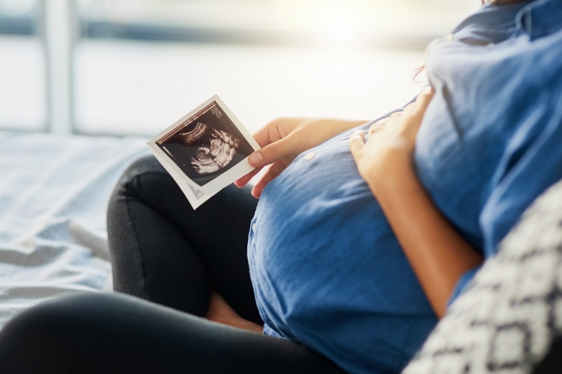
The differential diagnosis of a fetal lung mass includes congenital cystic adenomatoid malformation (CCAM), bronchopulmonary sequestration (BPS), hybrid lesions (CCAM and BPS elements), as well as other conditions such as bronchial atresia, bronchogenic cyst, esophageal duplication cyst, lobar emphysema and congenital high airway obstruction syndrome (CHAOS). These intrathoracic lesions can be confused with one another, but have distinct differences that require evaluation by a skilled team. For example, a right microcystic CCAM can appear to be a right congenital diaphragmatic hernia, while a BPS with a systemic arterial feeding vessel may also present as a solid chest mass, but cysts on ultrasound would make it more consistent with a hybrid lesion.

At CHOP, the experienced multidisciplinary team involved in the diagnosis and care of lung lesions — including high-risk obstetricians, pediatric surgeons, advanced practice nurses, radiologists, a coordinator, genetic counselors, social workers, anesthesiologists, neonatologists and cardiologists — is centrally located under one roof. Families that come to our center undergo a one-day comprehensive evaluation that includes Level II ultrasound using color flow Doppler, fetal echocardiogram and ultrafast fetal MRI, a technique we helped develop that can be very helpful in ruling out alternate diagnoses such as bronchial atresia, bronchogenic cyst or multilobar involvement.

Prenatal management and prognosis

Prenatal management and the overall prognosis for a patient with a lung lesion depends on the size and growth of the mass. More than 15 years ago, our imaging specialists developed the CCAM volume ratio (CVR) as a prognostic tool to measure the size of lung lesions before birth. This ratio helps determine the frequency of sonographic evaluation required over the duration of the pregnancy. Historically, we did not have a CVR to reassure families that their baby would not develop hydrops. Now, in many cases, we are able to predict the likelihood of hydrops developing.

Most lesions are followed by ultrasound and treated electively after birth. Esophageal compression by a large thoracic mass can interfere with fetal swallowing of amniotic fluid and result in polyhydramnios and potential preterm delivery. If a lesion continues to enlarge, further mediastinal shift occurs, resulting in compression of the heart and vena cava, which may lead to hydrops. Maternal mirror syndrome, a preeclamptic state where the mother mirrors the condition of her sick fetus, can occur in this scenario and requires careful maternal observation. When maternal mirror syndrome occurs, the only therapeutic alternative is delivery of the fetus, and invariably the sick, hydropic premature neonate dies.

For fetuses that have macrocystic CCAMs and hydrops, a fetal thoracoamniotic shunt can be employed to decompress the cyst fluid into the amniotic space and decrease the mass effect in the chest. For fetuses with solid lesions with a CVR > 1.6, maternal betamethasone can abrogate lesion growth. If betamethasone fails to stop the lesion’s growth and hydrops develops or persists, fetal resection is an option offered to families.

Our pediatric surgeons developed the open fetal surgical technique used to resect lung lesions. Intraoperative management of the mother and fetus is intensive. Some of the critical elements needed to carry out this procedure are sterile intraoperative ultrasound to determine the placental and fetal position; real-time fetal echocardiography,  provided by fetal cardiologists from our Fetal Heart Program, to determine cardiac function, aortic reversal of flow, ductal constriction, tricuspid regurgitation and decrease in fetal heart rate; employment of a miniaturized fetal pulse oximeter; and placement of an IV to deliver blood or medications.

Over the course of the pregnancy, our team works with referring physicians to determine the delivery location that will ensure the best outcome for mother and baby. Babies with small lesions can be delivered vaginally or via cesarean section (for obstetric indications) at a hospital close to home. Those with a mediastinal shift, but without hydrops or major lung compression, will likely be symptomatic and should be delivered by planned cesarean section at a quaternary center such as our Garbose Family Special Delivery Unit. This unit was specifically created to allow immediate access to the baby by necessary subspecialists and avoid transport and separation of mom from her newborn. Two operating rooms separated by a neonatal resuscitation room provide access to the baby for immediate resection of the lung lesion if necessary.

For those fetuses late in gestation with a large mediastinal shift and lung compression, we anticipate difficulties with neonatal ventilation and recommend ex utero intrapartum therapy (EXIT) delivery. The EXIT ensures uteroplacental gas exchange and fetal hemodynamic stability, converting a possible catastrophic emergency into a controlled, planned operation for mother and baby. We have performed more than 100 EXIT procedures for a variety of indications, by far the largest experience in the world. The  procedure requires a multidisciplinary team; deep general anesthesia for good uterine relaxation; supplemental fetal anesthesia by intramuscular vecuronium and fentanyl; and sterile intraoperative sonography.

Babies born asymptomatic are seen at 1 to 2 months of age by the pediatric surgeon. Typically a CT scan with IV contrast and the low-dose pediatric radiation protocol pioneered at CHOP is performed and these images are compared to the prenatal ultrasound and ultrafast fetal MRI. We offer postnatal resections by either a short muscle-sparing thoracotomy or thoracoscopy depending upon the presentation. We recommend elective resection between 1 to 3 months of age due to risks of infection and  malignant degeneration. Infants are usually discharged within 48 hours of the procedure, and follow-up occurs one month after surgery and then six months to one year later.

After the lung lesion is removed — whether before or after birth — the remaining normal lung tissue shows remarkable compensatory growth to fill the chest in children until about age 8. Early resection maximizes this compensatory lung growth and long-term follow-up has shown no residual respiratory problems as a result. Results with postnatal resection are excellent and parents should be reassured. We have come a long way in our understanding of lung lesions, but there is much more work to do. The  pathophysiology is complex and our Center for Fetal Research laboratory is focused on furthering our knowledge of these rare conditions.

References

Adzick NS. Management of fetal lung lesions. Clin Perinatol. 2009;36(2):363-376.

Peranteau WH, Wilson RD, Liechty KW, Johnson MP, Bebbington MW, Hedrick HL, Flake AW, Adzick NS. Effect of maternal betamethasone administration on prenatal congenital cystic adenomatoid malformation growth and fetal survival. Fetal Diagn Ther. 2007;22(5):365-371.

Hubbard AM, Adzick NS, Crombleholme TM, Coleman BG, Howell LJ, Haselgrove JC, Mahboubi S. Congenital chest lesions: diagnosis and characterization with prenatal MR imaging. Radiology. 1999 Jul;212(1):43-48.